Chlorine »
PDB 5u72-5ufe »
5udp »
Chlorine in PDB 5udp: High Resolution X-Ray Crystal Structure of Synthetic Insulin Lispro
Protein crystallography data
The structure of High Resolution X-Ray Crystal Structure of Synthetic Insulin Lispro, PDB code: 5udp
was solved by
K.Mandal,
B.Dhayalan,
S.B.H.Kent,
with X-Ray Crystallography technique. A brief refinement statistics is given in the table below:
| Resolution Low / High (Å) | 25.72 / 1.35 |
| Space group | P 1 21 1 |
| Cell size a, b, c (Å), α, β, γ (°) | 46.644, 61.470, 59.148, 90.00, 110.60, 90.00 |
| R / Rfree (%) | 16 / 19.4 |
Other elements in 5udp:
The structure of High Resolution X-Ray Crystal Structure of Synthetic Insulin Lispro also contains other interesting chemical elements:
| Zinc | (Zn) | 2 atoms |
| Sodium | (Na) | 1 atom |
Chlorine Binding Sites:
The binding sites of Chlorine atom in the High Resolution X-Ray Crystal Structure of Synthetic Insulin Lispro
(pdb code 5udp). This binding sites where shown within
5.0 Angstroms radius around Chlorine atom.
In total 2 binding sites of Chlorine where determined in the High Resolution X-Ray Crystal Structure of Synthetic Insulin Lispro, PDB code: 5udp:
Jump to Chlorine binding site number: 1; 2;
In total 2 binding sites of Chlorine where determined in the High Resolution X-Ray Crystal Structure of Synthetic Insulin Lispro, PDB code: 5udp:
Jump to Chlorine binding site number: 1; 2;
Chlorine binding site 1 out of 2 in 5udp
Go back to
Chlorine binding site 1 out
of 2 in the High Resolution X-Ray Crystal Structure of Synthetic Insulin Lispro
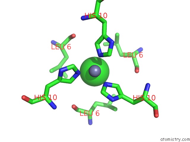
Mono view
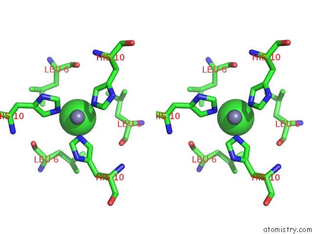
Stereo pair view
Mono view
Stereo pair view
A full contact list of Chlorine with other atoms in the Cl binding
site number 1 of High Resolution X-Ray Crystal Structure of Synthetic Insulin Lispro within 5.0Å range:
|
Chlorine binding site 2 out of 2 in 5udp
Go back to
Chlorine binding site 2 out
of 2 in the High Resolution X-Ray Crystal Structure of Synthetic Insulin Lispro
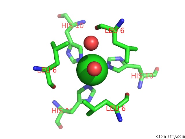
Mono view
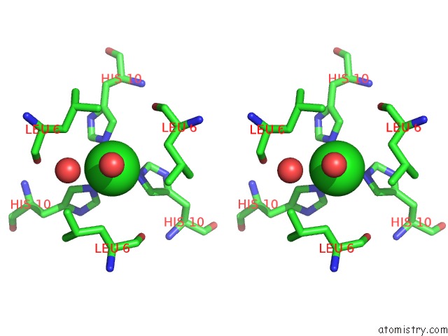
Stereo pair view
Mono view
Stereo pair view
A full contact list of Chlorine with other atoms in the Cl binding
site number 2 of High Resolution X-Ray Crystal Structure of Synthetic Insulin Lispro within 5.0Å range:
|
Reference:
B.Dhayalan,
K.Mandal,
N.Rege,
M.A.Weiss,
S.H.Eitel,
T.Meier,
R.O.Schoenleber,
S.B.Kent.
Scope and Limitations of Fmoc Chemistry Spps-Based Approaches to the Total Synthesis of Insulin Lispro Via Ester Insulin. Chemistry V. 23 1709 2017.
ISSN: ISSN 1521-3765
PubMed: 27905149
DOI: 10.1002/CHEM.201605578
Page generated: Fri Jul 26 18:02:27 2024
ISSN: ISSN 1521-3765
PubMed: 27905149
DOI: 10.1002/CHEM.201605578
Last articles
Zn in 9J0NZn in 9J0O
Zn in 9J0P
Zn in 9FJX
Zn in 9EKB
Zn in 9C0F
Zn in 9CAH
Zn in 9CH0
Zn in 9CH3
Zn in 9CH1