Chlorine »
PDB 5uwx-5v4g »
5v36 »
Chlorine in PDB 5v36: 1.88 Angstrom Resolution Crystal Structure of Glutathione Reductase From Streptococcus Mutans UA159 in Complex with Fad
Enzymatic activity of 1.88 Angstrom Resolution Crystal Structure of Glutathione Reductase From Streptococcus Mutans UA159 in Complex with Fad
All present enzymatic activity of 1.88 Angstrom Resolution Crystal Structure of Glutathione Reductase From Streptococcus Mutans UA159 in Complex with Fad:
1.8.1.7; 3.3.1.1;
1.8.1.7; 3.3.1.1;
Protein crystallography data
The structure of 1.88 Angstrom Resolution Crystal Structure of Glutathione Reductase From Streptococcus Mutans UA159 in Complex with Fad, PDB code: 5v36
was solved by
G.Minasov,
L.Shuvalova,
I.Dubrovska,
O.Kiryukhina,
S.Grimshaw,
K.Kwon,
W.F.Anderson,
Center For Structural Genomics Of Infectious Diseases(Csgid),
with X-Ray Crystallography technique. A brief refinement statistics is given in the table below:
| Resolution Low / High (Å) | 29.81 / 1.88 |
| Space group | P 65 2 2 |
| Cell size a, b, c (Å), α, β, γ (°) | 126.333, 126.333, 247.876, 90.00, 90.00, 120.00 |
| R / Rfree (%) | 14.3 / 17 |
Chlorine Binding Sites:
The binding sites of Chlorine atom in the 1.88 Angstrom Resolution Crystal Structure of Glutathione Reductase From Streptococcus Mutans UA159 in Complex with Fad
(pdb code 5v36). This binding sites where shown within
5.0 Angstroms radius around Chlorine atom.
In total 10 binding sites of Chlorine where determined in the 1.88 Angstrom Resolution Crystal Structure of Glutathione Reductase From Streptococcus Mutans UA159 in Complex with Fad, PDB code: 5v36:
Jump to Chlorine binding site number: 1; 2; 3; 4; 5; 6; 7; 8; 9; 10;
In total 10 binding sites of Chlorine where determined in the 1.88 Angstrom Resolution Crystal Structure of Glutathione Reductase From Streptococcus Mutans UA159 in Complex with Fad, PDB code: 5v36:
Jump to Chlorine binding site number: 1; 2; 3; 4; 5; 6; 7; 8; 9; 10;
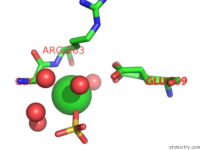
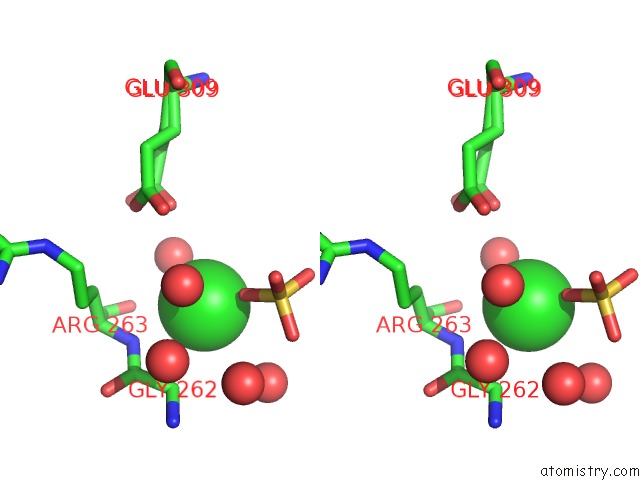
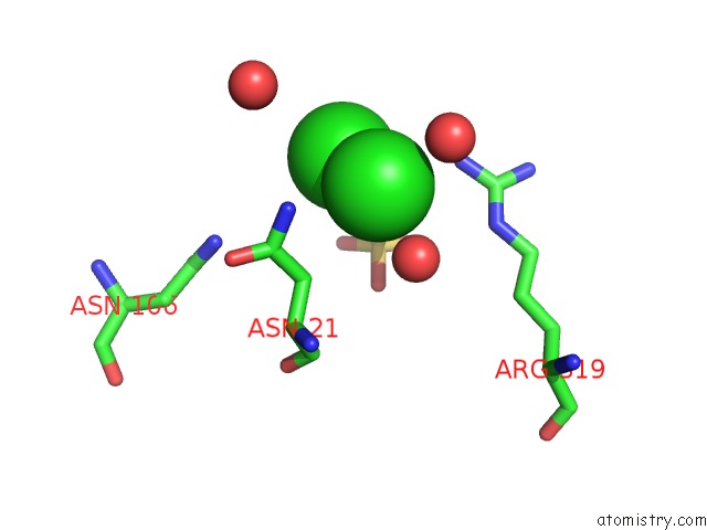
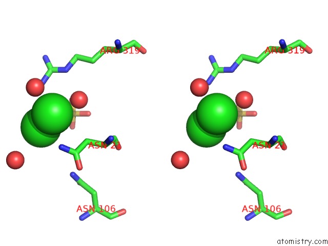
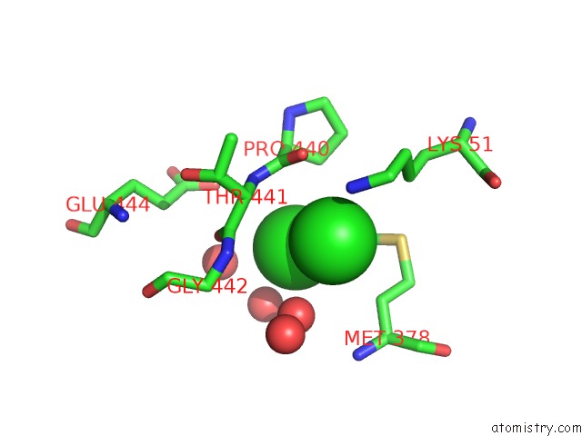
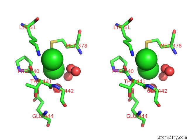
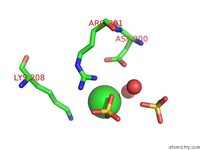
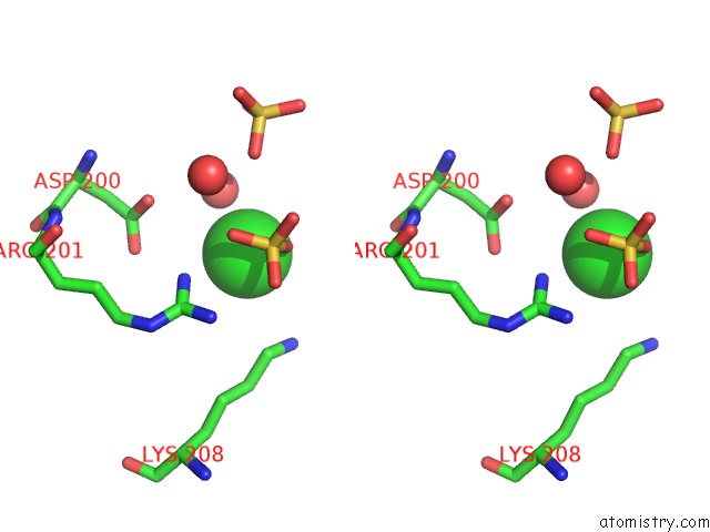
Chlorine binding site 1 out of 10 in 5v36
Go back to
Chlorine binding site 1 out
of 10 in the 1.88 Angstrom Resolution Crystal Structure of Glutathione Reductase From Streptococcus Mutans UA159 in Complex with Fad
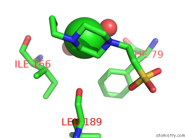
Mono view
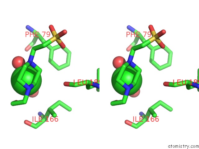
Stereo pair view
Mono view
Stereo pair view
A full contact list of Chlorine with other atoms in the Cl binding
site number 1 of 1.88 Angstrom Resolution Crystal Structure of Glutathione Reductase From Streptococcus Mutans UA159 in Complex with Fad within 5.0Å range:
|
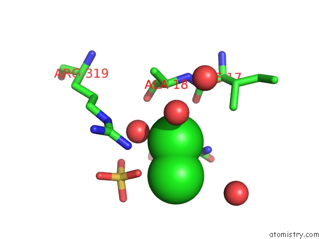
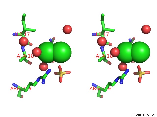
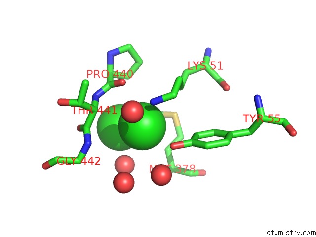
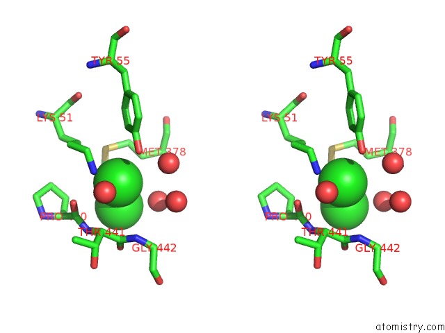
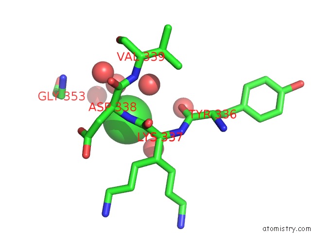
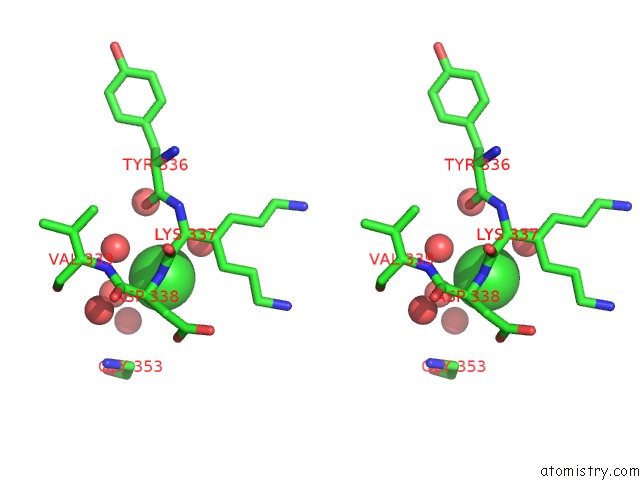
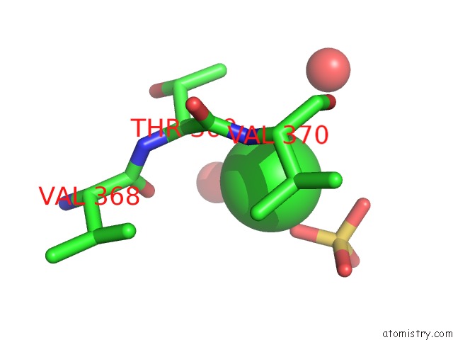
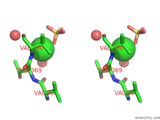
Chlorine binding site 2 out of 10 in 5v36
Go back to
Chlorine binding site 2 out
of 10 in the 1.88 Angstrom Resolution Crystal Structure of Glutathione Reductase From Streptococcus Mutans UA159 in Complex with Fad
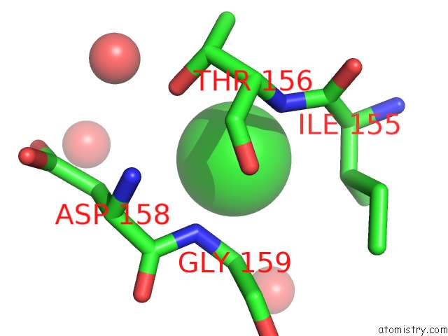
Mono view
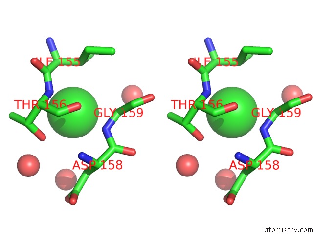
Stereo pair view
Mono view
Stereo pair view
A full contact list of Chlorine with other atoms in the Cl binding
site number 2 of 1.88 Angstrom Resolution Crystal Structure of Glutathione Reductase From Streptococcus Mutans UA159 in Complex with Fad within 5.0Å range:
|
Chlorine binding site 3 out of 10 in 5v36
Go back to
Chlorine binding site 3 out
of 10 in the 1.88 Angstrom Resolution Crystal Structure of Glutathione Reductase From Streptococcus Mutans UA159 in Complex with Fad
Mono view
Stereo pair view
Mono view
Stereo pair view
A full contact list of Chlorine with other atoms in the Cl binding
site number 3 of 1.88 Angstrom Resolution Crystal Structure of Glutathione Reductase From Streptococcus Mutans UA159 in Complex with Fad within 5.0Å range:
|
Chlorine binding site 4 out of 10 in 5v36
Go back to
Chlorine binding site 4 out
of 10 in the 1.88 Angstrom Resolution Crystal Structure of Glutathione Reductase From Streptococcus Mutans UA159 in Complex with Fad
Mono view
Stereo pair view
Mono view
Stereo pair view
A full contact list of Chlorine with other atoms in the Cl binding
site number 4 of 1.88 Angstrom Resolution Crystal Structure of Glutathione Reductase From Streptococcus Mutans UA159 in Complex with Fad within 5.0Å range:
|
Chlorine binding site 5 out of 10 in 5v36
Go back to
Chlorine binding site 5 out
of 10 in the 1.88 Angstrom Resolution Crystal Structure of Glutathione Reductase From Streptococcus Mutans UA159 in Complex with Fad
Mono view
Stereo pair view
Mono view
Stereo pair view
A full contact list of Chlorine with other atoms in the Cl binding
site number 5 of 1.88 Angstrom Resolution Crystal Structure of Glutathione Reductase From Streptococcus Mutans UA159 in Complex with Fad within 5.0Å range:
|
Chlorine binding site 6 out of 10 in 5v36
Go back to
Chlorine binding site 6 out
of 10 in the 1.88 Angstrom Resolution Crystal Structure of Glutathione Reductase From Streptococcus Mutans UA159 in Complex with Fad
Mono view
Stereo pair view
Mono view
Stereo pair view
A full contact list of Chlorine with other atoms in the Cl binding
site number 6 of 1.88 Angstrom Resolution Crystal Structure of Glutathione Reductase From Streptococcus Mutans UA159 in Complex with Fad within 5.0Å range:
|
Chlorine binding site 7 out of 10 in 5v36
Go back to
Chlorine binding site 7 out
of 10 in the 1.88 Angstrom Resolution Crystal Structure of Glutathione Reductase From Streptococcus Mutans UA159 in Complex with Fad
Mono view
Stereo pair view
Mono view
Stereo pair view
A full contact list of Chlorine with other atoms in the Cl binding
site number 7 of 1.88 Angstrom Resolution Crystal Structure of Glutathione Reductase From Streptococcus Mutans UA159 in Complex with Fad within 5.0Å range:
|
Chlorine binding site 8 out of 10 in 5v36
Go back to
Chlorine binding site 8 out
of 10 in the 1.88 Angstrom Resolution Crystal Structure of Glutathione Reductase From Streptococcus Mutans UA159 in Complex with Fad
Mono view
Stereo pair view
Mono view
Stereo pair view
A full contact list of Chlorine with other atoms in the Cl binding
site number 8 of 1.88 Angstrom Resolution Crystal Structure of Glutathione Reductase From Streptococcus Mutans UA159 in Complex with Fad within 5.0Å range:
|
Chlorine binding site 9 out of 10 in 5v36
Go back to
Chlorine binding site 9 out
of 10 in the 1.88 Angstrom Resolution Crystal Structure of Glutathione Reductase From Streptococcus Mutans UA159 in Complex with Fad
Mono view
Stereo pair view
Mono view
Stereo pair view
A full contact list of Chlorine with other atoms in the Cl binding
site number 9 of 1.88 Angstrom Resolution Crystal Structure of Glutathione Reductase From Streptococcus Mutans UA159 in Complex with Fad within 5.0Å range:
|
Chlorine binding site 10 out of 10 in 5v36
Go back to
Chlorine binding site 10 out
of 10 in the 1.88 Angstrom Resolution Crystal Structure of Glutathione Reductase From Streptococcus Mutans UA159 in Complex with Fad
Mono view
Stereo pair view
Mono view
Stereo pair view
A full contact list of Chlorine with other atoms in the Cl binding
site number 10 of 1.88 Angstrom Resolution Crystal Structure of Glutathione Reductase From Streptococcus Mutans UA159 in Complex with Fad within 5.0Å range:
|
Reference:
G.Minasov,
L.Shuvalova,
I.Dubrovska,
O.Kiryukhina,
S.Grimshaw,
K.Kwon,
W.F.Anderson,
Center For Structural Genomics Of Infectious Diseases(Csgid).
1.88 Angstrom Resolution Crystal Structure of Glutathione Reductase From Streptococcus Mutans UA159 in Complex with Fad. To Be Published.
Page generated: Sat Jul 12 09:42:10 2025
Last articles
Cl in 8CW0Cl in 8CVV
Cl in 8CVU
Cl in 8CUR
Cl in 8CU9
Cl in 8CTY
Cl in 8CTM
Cl in 8CSD
Cl in 8CP8
Cl in 8CTB