Chlorine »
PDB 5vc5-5vm6 »
5vcy »
Chlorine in PDB 5vcy: Crystal Structure of Human MYT1 Kinase Domain in Complex with Bosutinib
Enzymatic activity of Crystal Structure of Human MYT1 Kinase Domain in Complex with Bosutinib
All present enzymatic activity of Crystal Structure of Human MYT1 Kinase Domain in Complex with Bosutinib:
2.7.11.1;
2.7.11.1;
Protein crystallography data
The structure of Crystal Structure of Human MYT1 Kinase Domain in Complex with Bosutinib, PDB code: 5vcy
was solved by
J.-Y.Zhu,
E.Schonbrunn,
with X-Ray Crystallography technique. A brief refinement statistics is given in the table below:
| Resolution Low / High (Å) | 36.05 / 1.56 |
| Space group | P 21 21 21 |
| Cell size a, b, c (Å), α, β, γ (°) | 46.630, 55.000, 113.660, 90.00, 90.00, 90.00 |
| R / Rfree (%) | 14 / 17.8 |
Chlorine Binding Sites:
The binding sites of Chlorine atom in the Crystal Structure of Human MYT1 Kinase Domain in Complex with Bosutinib
(pdb code 5vcy). This binding sites where shown within
5.0 Angstroms radius around Chlorine atom.
In total 2 binding sites of Chlorine where determined in the Crystal Structure of Human MYT1 Kinase Domain in Complex with Bosutinib, PDB code: 5vcy:
Jump to Chlorine binding site number: 1; 2;
In total 2 binding sites of Chlorine where determined in the Crystal Structure of Human MYT1 Kinase Domain in Complex with Bosutinib, PDB code: 5vcy:
Jump to Chlorine binding site number: 1; 2;
Chlorine binding site 1 out of 2 in 5vcy
Go back to
Chlorine binding site 1 out
of 2 in the Crystal Structure of Human MYT1 Kinase Domain in Complex with Bosutinib
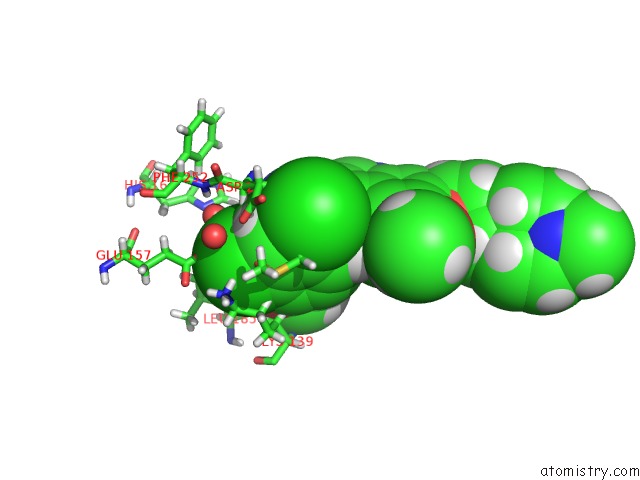
Mono view

Stereo pair view
Mono view
Stereo pair view
A full contact list of Chlorine with other atoms in the Cl binding
site number 1 of Crystal Structure of Human MYT1 Kinase Domain in Complex with Bosutinib within 5.0Å range:
|
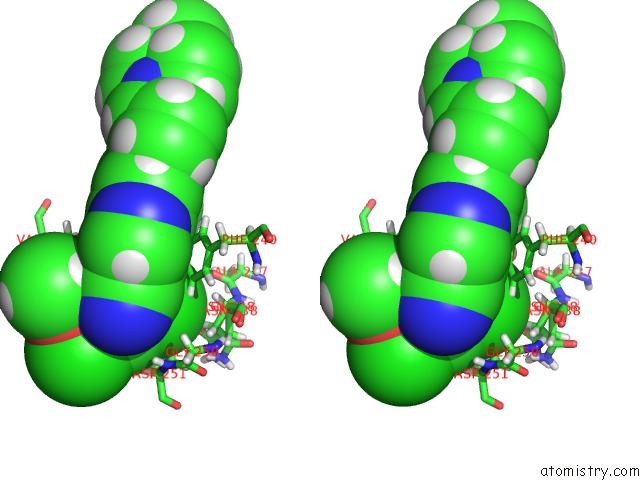
Chlorine binding site 2 out of 2 in 5vcy
Go back to
Chlorine binding site 2 out
of 2 in the Crystal Structure of Human MYT1 Kinase Domain in Complex with Bosutinib

Mono view
Stereo pair view
Mono view
Stereo pair view
A full contact list of Chlorine with other atoms in the Cl binding
site number 2 of Crystal Structure of Human MYT1 Kinase Domain in Complex with Bosutinib within 5.0Å range:
|
Reference:
J.Y.Zhu,
R.A.Cuellar,
N.Berndt,
H.E.Lee,
S.H.Olesen,
M.P.Martin,
J.T.Jensen,
G.I.Georg,
E.Schonbrunn.
Structural Basis of Wee Kinases Functionality and Inactivation By Diverse Small Molecule Inhibitors. J. Med. Chem. V. 60 7863 2017.
ISSN: ISSN 1520-4804
PubMed: 28792760
DOI: 10.1021/ACS.JMEDCHEM.7B00996
Page generated: Sat Jul 12 09:50:41 2025
ISSN: ISSN 1520-4804
PubMed: 28792760
DOI: 10.1021/ACS.JMEDCHEM.7B00996
Last articles
Cl in 6GGICl in 6GGL
Cl in 6GGK
Cl in 6GES
Cl in 6GGG
Cl in 6GFS
Cl in 6GEO
Cl in 6GDQ
Cl in 6GE0
Cl in 6GCW