Chlorine »
PDB 5vmq-5vtk »
5vqb »
Chlorine in PDB 5vqb: Crystal Structure of Rifampin Monooxygenase From Streptomyces Venezuelae, Complex with Fad
Protein crystallography data
The structure of Crystal Structure of Rifampin Monooxygenase From Streptomyces Venezuelae, Complex with Fad, PDB code: 5vqb
was solved by
G.Cox,
J.Kelso,
P.J.Stogios,
A.Savchenko,
W.F.Anderson,
G.D.Wright,
Centerfor Structural Genomics Of Infectious Diseases (Csgid),
with X-Ray Crystallography technique. A brief refinement statistics is given in the table below:
| Resolution Low / High (Å) | 48.92 / 3.39 |
| Space group | C 1 2 1 |
| Cell size a, b, c (Å), α, β, γ (°) | 203.085, 129.285, 75.311, 90.00, 105.52, 90.00 |
| R / Rfree (%) | 22.6 / 28.4 |
Chlorine Binding Sites:
The binding sites of Chlorine atom in the Crystal Structure of Rifampin Monooxygenase From Streptomyces Venezuelae, Complex with Fad
(pdb code 5vqb). This binding sites where shown within
5.0 Angstroms radius around Chlorine atom.
In total 8 binding sites of Chlorine where determined in the Crystal Structure of Rifampin Monooxygenase From Streptomyces Venezuelae, Complex with Fad, PDB code: 5vqb:
Jump to Chlorine binding site number: 1; 2; 3; 4; 5; 6; 7; 8;
In total 8 binding sites of Chlorine where determined in the Crystal Structure of Rifampin Monooxygenase From Streptomyces Venezuelae, Complex with Fad, PDB code: 5vqb:
Jump to Chlorine binding site number: 1; 2; 3; 4; 5; 6; 7; 8;
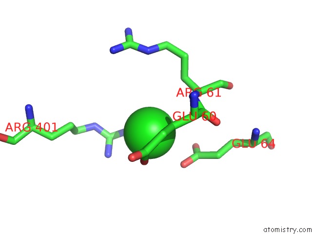
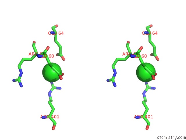
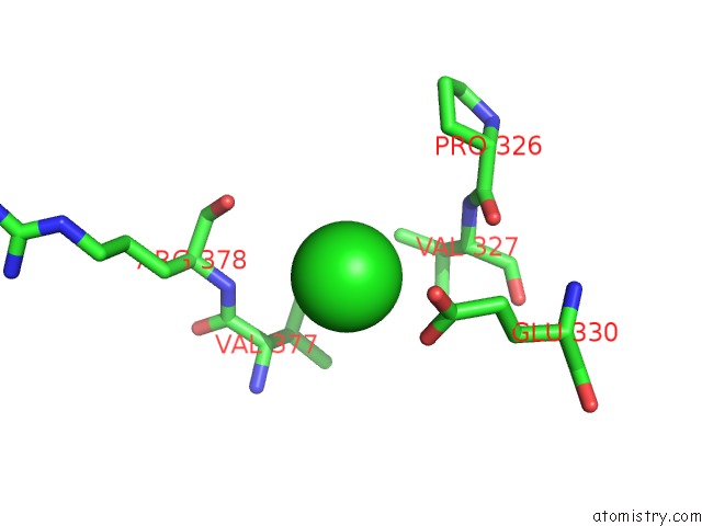
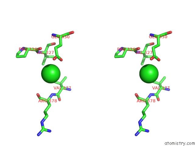
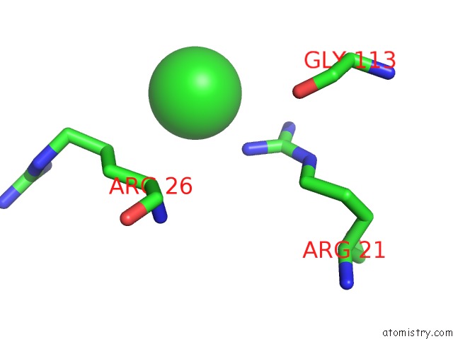
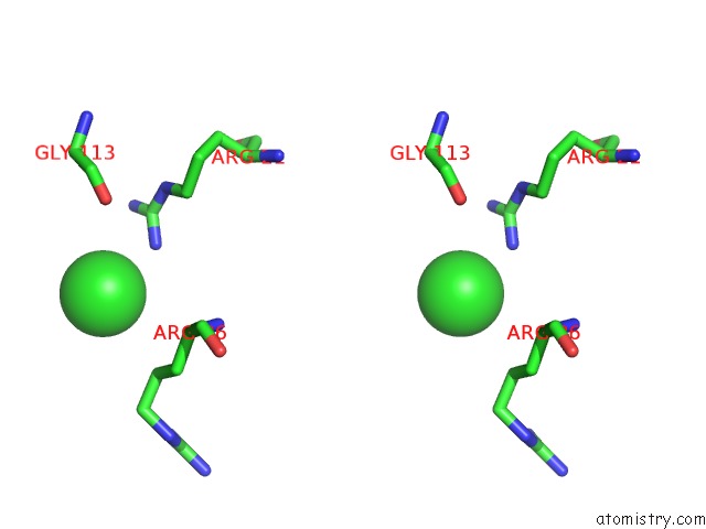
Chlorine binding site 1 out of 8 in 5vqb
Go back to
Chlorine binding site 1 out
of 8 in the Crystal Structure of Rifampin Monooxygenase From Streptomyces Venezuelae, Complex with Fad
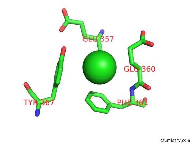
Mono view
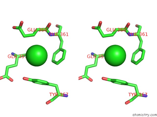
Stereo pair view
Mono view
Stereo pair view
A full contact list of Chlorine with other atoms in the Cl binding
site number 1 of Crystal Structure of Rifampin Monooxygenase From Streptomyces Venezuelae, Complex with Fad within 5.0Å range:
|
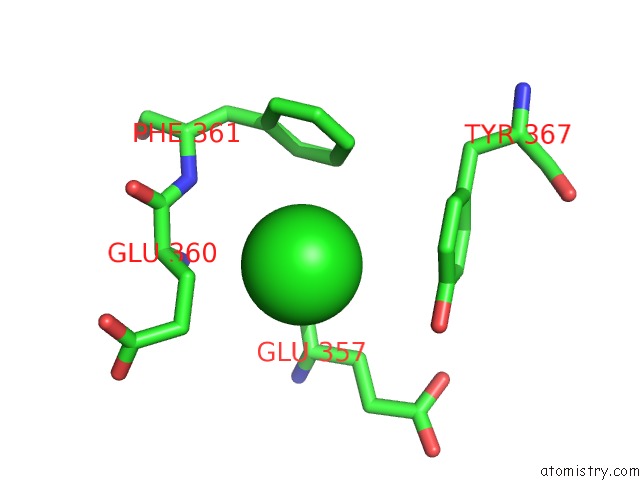
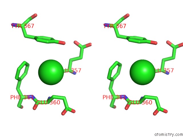
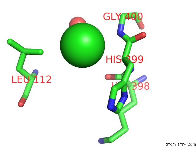
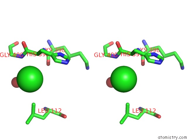
Chlorine binding site 2 out of 8 in 5vqb
Go back to
Chlorine binding site 2 out
of 8 in the Crystal Structure of Rifampin Monooxygenase From Streptomyces Venezuelae, Complex with Fad
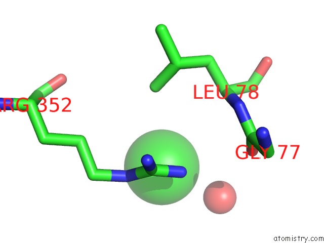
Mono view
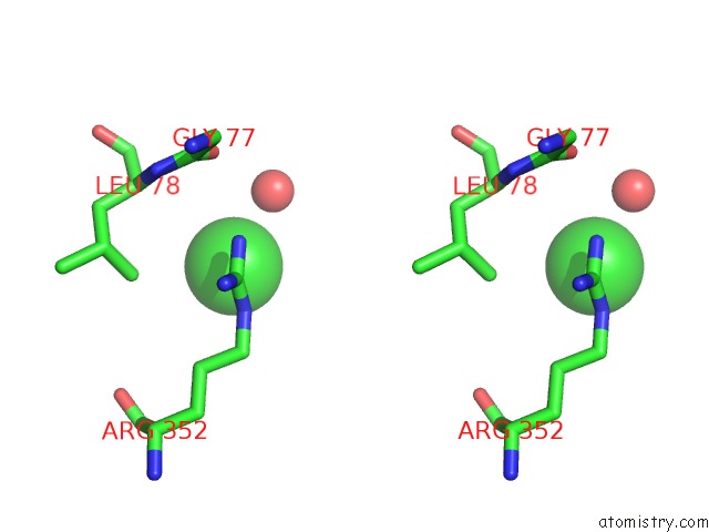
Stereo pair view
Mono view
Stereo pair view
A full contact list of Chlorine with other atoms in the Cl binding
site number 2 of Crystal Structure of Rifampin Monooxygenase From Streptomyces Venezuelae, Complex with Fad within 5.0Å range:
|
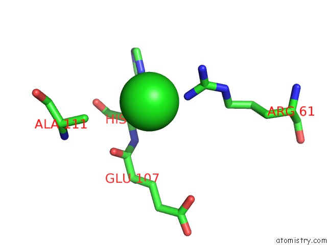
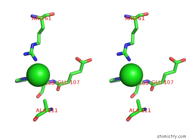
Chlorine binding site 3 out of 8 in 5vqb
Go back to
Chlorine binding site 3 out
of 8 in the Crystal Structure of Rifampin Monooxygenase From Streptomyces Venezuelae, Complex with Fad
Mono view
Stereo pair view
Mono view
Stereo pair view
A full contact list of Chlorine with other atoms in the Cl binding
site number 3 of Crystal Structure of Rifampin Monooxygenase From Streptomyces Venezuelae, Complex with Fad within 5.0Å range:
|
Chlorine binding site 4 out of 8 in 5vqb
Go back to
Chlorine binding site 4 out
of 8 in the Crystal Structure of Rifampin Monooxygenase From Streptomyces Venezuelae, Complex with Fad
Mono view
Stereo pair view
Mono view
Stereo pair view
A full contact list of Chlorine with other atoms in the Cl binding
site number 4 of Crystal Structure of Rifampin Monooxygenase From Streptomyces Venezuelae, Complex with Fad within 5.0Å range:
|
Chlorine binding site 5 out of 8 in 5vqb
Go back to
Chlorine binding site 5 out
of 8 in the Crystal Structure of Rifampin Monooxygenase From Streptomyces Venezuelae, Complex with Fad
Mono view
Stereo pair view
Mono view
Stereo pair view
A full contact list of Chlorine with other atoms in the Cl binding
site number 5 of Crystal Structure of Rifampin Monooxygenase From Streptomyces Venezuelae, Complex with Fad within 5.0Å range:
|
Chlorine binding site 6 out of 8 in 5vqb
Go back to
Chlorine binding site 6 out
of 8 in the Crystal Structure of Rifampin Monooxygenase From Streptomyces Venezuelae, Complex with Fad
Mono view
Stereo pair view
Mono view
Stereo pair view
A full contact list of Chlorine with other atoms in the Cl binding
site number 6 of Crystal Structure of Rifampin Monooxygenase From Streptomyces Venezuelae, Complex with Fad within 5.0Å range:
|
Chlorine binding site 7 out of 8 in 5vqb
Go back to
Chlorine binding site 7 out
of 8 in the Crystal Structure of Rifampin Monooxygenase From Streptomyces Venezuelae, Complex with Fad
Mono view
Stereo pair view
Mono view
Stereo pair view
A full contact list of Chlorine with other atoms in the Cl binding
site number 7 of Crystal Structure of Rifampin Monooxygenase From Streptomyces Venezuelae, Complex with Fad within 5.0Å range:
|
Chlorine binding site 8 out of 8 in 5vqb
Go back to
Chlorine binding site 8 out
of 8 in the Crystal Structure of Rifampin Monooxygenase From Streptomyces Venezuelae, Complex with Fad
Mono view
Stereo pair view
Mono view
Stereo pair view
A full contact list of Chlorine with other atoms in the Cl binding
site number 8 of Crystal Structure of Rifampin Monooxygenase From Streptomyces Venezuelae, Complex with Fad within 5.0Å range:
|
Reference:
K.Koteva,
G.Cox,
J.K.Kelso,
M.D.Surette,
H.L.Zubyk,
L.Ejim,
P.Stogios,
A.Savchenko,
D.Sorensen,
G.D.Wright.
Rox, A Rifamycin Resistance Enzyme with An Unprecedented Mechanism of Action. Cell Chem Biol V. 25 403 2018.
ISSN: ESSN 2451-9448
PubMed: 29398560
DOI: 10.1016/J.CHEMBIOL.2018.01.009
Page generated: Sat Jul 12 09:59:44 2025
ISSN: ESSN 2451-9448
PubMed: 29398560
DOI: 10.1016/J.CHEMBIOL.2018.01.009
Last articles
F in 4FM7F in 4FLH
F in 4FIA
F in 4FKI
F in 4FK3
F in 4FJZ
F in 4FJY
F in 4FF6
F in 4FIM
F in 4FDO