Chlorine »
PDB 5w5o-5we8 »
5w6s »
Chlorine in PDB 5w6s: Crystal Structure of Bacteriophage CBA120 Tailspike Protein 2 Enzymatically Active Domain (TSP2DN, ORF211) Complex with Escherichia Coli O157-Antigen
Protein crystallography data
The structure of Crystal Structure of Bacteriophage CBA120 Tailspike Protein 2 Enzymatically Active Domain (TSP2DN, ORF211) Complex with Escherichia Coli O157-Antigen, PDB code: 5w6s
was solved by
M.Plattner,
M.M.Shneider,
P.G.Leiman,
with X-Ray Crystallography technique. A brief refinement statistics is given in the table below:
| Resolution Low / High (Å) | 49.50 / 2.26 |
| Space group | P 43 3 2 |
| Cell size a, b, c (Å), α, β, γ (°) | 185.227, 185.227, 185.227, 90.00, 90.00, 90.00 |
| R / Rfree (%) | 13.3 / 16.4 |
Other elements in 5w6s:
The structure of Crystal Structure of Bacteriophage CBA120 Tailspike Protein 2 Enzymatically Active Domain (TSP2DN, ORF211) Complex with Escherichia Coli O157-Antigen also contains other interesting chemical elements:
| Potassium | (K) | 1 atom |
| Sodium | (Na) | 1 atom |
Chlorine Binding Sites:
The binding sites of Chlorine atom in the Crystal Structure of Bacteriophage CBA120 Tailspike Protein 2 Enzymatically Active Domain (TSP2DN, ORF211) Complex with Escherichia Coli O157-Antigen
(pdb code 5w6s). This binding sites where shown within
5.0 Angstroms radius around Chlorine atom.
In total 3 binding sites of Chlorine where determined in the Crystal Structure of Bacteriophage CBA120 Tailspike Protein 2 Enzymatically Active Domain (TSP2DN, ORF211) Complex with Escherichia Coli O157-Antigen, PDB code: 5w6s:
Jump to Chlorine binding site number: 1; 2; 3;
In total 3 binding sites of Chlorine where determined in the Crystal Structure of Bacteriophage CBA120 Tailspike Protein 2 Enzymatically Active Domain (TSP2DN, ORF211) Complex with Escherichia Coli O157-Antigen, PDB code: 5w6s:
Jump to Chlorine binding site number: 1; 2; 3;
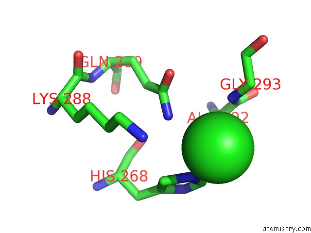
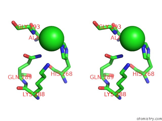
Chlorine binding site 1 out of 3 in 5w6s
Go back to
Chlorine binding site 1 out
of 3 in the Crystal Structure of Bacteriophage CBA120 Tailspike Protein 2 Enzymatically Active Domain (TSP2DN, ORF211) Complex with Escherichia Coli O157-Antigen
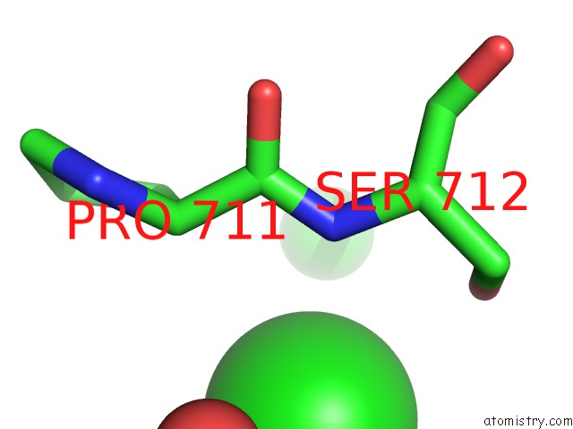
Mono view
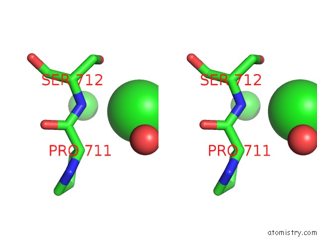
Stereo pair view
Mono view
Stereo pair view
A full contact list of Chlorine with other atoms in the Cl binding
site number 1 of Crystal Structure of Bacteriophage CBA120 Tailspike Protein 2 Enzymatically Active Domain (TSP2DN, ORF211) Complex with Escherichia Coli O157-Antigen within 5.0Å range:
|
Chlorine binding site 2 out of 3 in 5w6s
Go back to
Chlorine binding site 2 out
of 3 in the Crystal Structure of Bacteriophage CBA120 Tailspike Protein 2 Enzymatically Active Domain (TSP2DN, ORF211) Complex with Escherichia Coli O157-Antigen
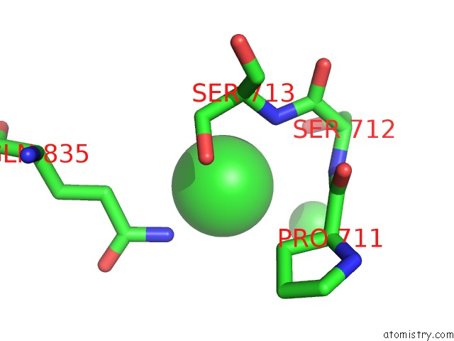
Mono view
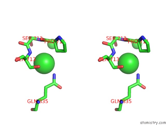
Stereo pair view
Mono view
Stereo pair view
A full contact list of Chlorine with other atoms in the Cl binding
site number 2 of Crystal Structure of Bacteriophage CBA120 Tailspike Protein 2 Enzymatically Active Domain (TSP2DN, ORF211) Complex with Escherichia Coli O157-Antigen within 5.0Å range:
|
Chlorine binding site 3 out of 3 in 5w6s
Go back to
Chlorine binding site 3 out
of 3 in the Crystal Structure of Bacteriophage CBA120 Tailspike Protein 2 Enzymatically Active Domain (TSP2DN, ORF211) Complex with Escherichia Coli O157-Antigen
Mono view
Stereo pair view
Mono view
Stereo pair view
A full contact list of Chlorine with other atoms in the Cl binding
site number 3 of Crystal Structure of Bacteriophage CBA120 Tailspike Protein 2 Enzymatically Active Domain (TSP2DN, ORF211) Complex with Escherichia Coli O157-Antigen within 5.0Å range:
|
Reference:
M.Plattner,
M.M.Shneider,
N.P.Arbatsky,
A.S.Shashkov,
A.O.Chizhov,
S.Nazarov,
N.S.Prokhorov,
N.M.I.Taylor,
S.A.Buth,
M.Gambino,
Y.E.Gencay,
L.Brondsted,
E.M.Kutter,
Y.A.Knirel,
P.G.Leiman.
Structure and Function of the Branched Receptor-Binding Complex of Bacteriophage CBA120. J.Mol.Biol. V. 431 3718 2019.
ISSN: ESSN 1089-8638
PubMed: 31325442
DOI: 10.1016/J.JMB.2019.07.022
Page generated: Fri Jul 26 19:13:47 2024
ISSN: ESSN 1089-8638
PubMed: 31325442
DOI: 10.1016/J.JMB.2019.07.022
Last articles
Zn in 9J0NZn in 9J0O
Zn in 9J0P
Zn in 9FJX
Zn in 9EKB
Zn in 9C0F
Zn in 9CAH
Zn in 9CH0
Zn in 9CH3
Zn in 9CH1