Chlorine »
PDB 5w5o-5we8 »
5wbg »
Chlorine in PDB 5wbg: Crystal Structure of Human Cytochrome P450 2B6 (Y226H/K262R) in Complex with An Analog of A Drug Efavirenz
Protein crystallography data
The structure of Crystal Structure of Human Cytochrome P450 2B6 (Y226H/K262R) in Complex with An Analog of A Drug Efavirenz, PDB code: 5wbg
was solved by
M.B.Shah,
J.R.Halpert,
with X-Ray Crystallography technique. A brief refinement statistics is given in the table below:
| Resolution Low / High (Å) | 50.00 / 2.99 |
| Space group | P 1 21 1 |
| Cell size a, b, c (Å), α, β, γ (°) | 103.293, 197.786, 119.222, 90.00, 98.51, 90.00 |
| R / Rfree (%) | 22.7 / 26.2 |
Other elements in 5wbg:
The structure of Crystal Structure of Human Cytochrome P450 2B6 (Y226H/K262R) in Complex with An Analog of A Drug Efavirenz also contains other interesting chemical elements:
| Fluorine | (F) | 18 atoms |
| Iron | (Fe) | 6 atoms |
Chlorine Binding Sites:
The binding sites of Chlorine atom in the Crystal Structure of Human Cytochrome P450 2B6 (Y226H/K262R) in Complex with An Analog of A Drug Efavirenz
(pdb code 5wbg). This binding sites where shown within
5.0 Angstroms radius around Chlorine atom.
In total 6 binding sites of Chlorine where determined in the Crystal Structure of Human Cytochrome P450 2B6 (Y226H/K262R) in Complex with An Analog of A Drug Efavirenz, PDB code: 5wbg:
Jump to Chlorine binding site number: 1; 2; 3; 4; 5; 6;
In total 6 binding sites of Chlorine where determined in the Crystal Structure of Human Cytochrome P450 2B6 (Y226H/K262R) in Complex with An Analog of A Drug Efavirenz, PDB code: 5wbg:
Jump to Chlorine binding site number: 1; 2; 3; 4; 5; 6;
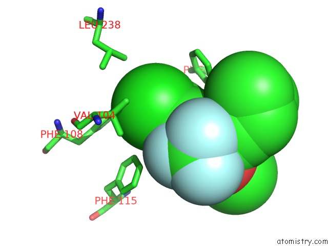
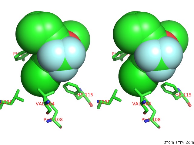
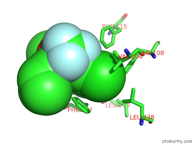
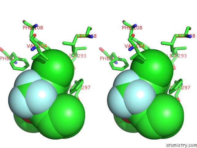
Chlorine binding site 1 out of 6 in 5wbg
Go back to
Chlorine binding site 1 out
of 6 in the Crystal Structure of Human Cytochrome P450 2B6 (Y226H/K262R) in Complex with An Analog of A Drug Efavirenz
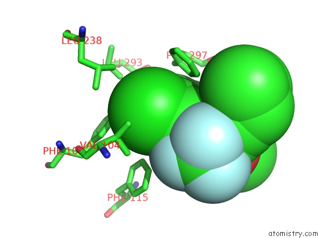
Mono view
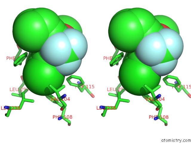
Stereo pair view
Mono view
Stereo pair view
A full contact list of Chlorine with other atoms in the Cl binding
site number 1 of Crystal Structure of Human Cytochrome P450 2B6 (Y226H/K262R) in Complex with An Analog of A Drug Efavirenz within 5.0Å range:
|
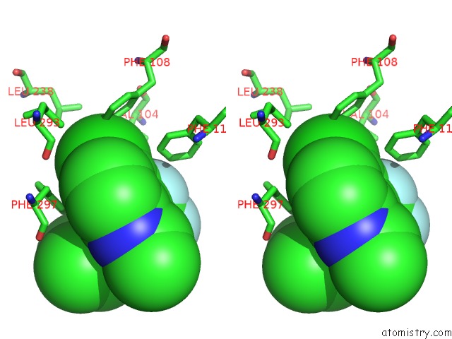
Chlorine binding site 2 out of 6 in 5wbg
Go back to
Chlorine binding site 2 out
of 6 in the Crystal Structure of Human Cytochrome P450 2B6 (Y226H/K262R) in Complex with An Analog of A Drug Efavirenz
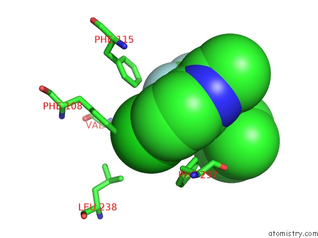
Mono view
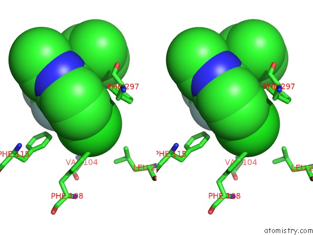
Stereo pair view
Mono view
Stereo pair view
A full contact list of Chlorine with other atoms in the Cl binding
site number 2 of Crystal Structure of Human Cytochrome P450 2B6 (Y226H/K262R) in Complex with An Analog of A Drug Efavirenz within 5.0Å range:
|
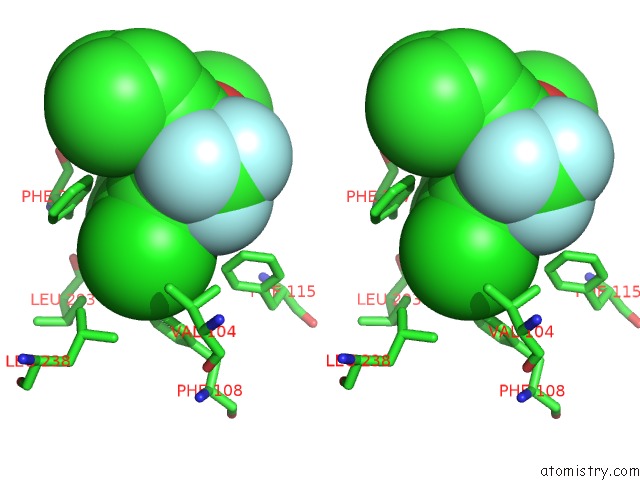
Chlorine binding site 3 out of 6 in 5wbg
Go back to
Chlorine binding site 3 out
of 6 in the Crystal Structure of Human Cytochrome P450 2B6 (Y226H/K262R) in Complex with An Analog of A Drug Efavirenz
Mono view
Stereo pair view
Mono view
Stereo pair view
A full contact list of Chlorine with other atoms in the Cl binding
site number 3 of Crystal Structure of Human Cytochrome P450 2B6 (Y226H/K262R) in Complex with An Analog of A Drug Efavirenz within 5.0Å range:
|
Chlorine binding site 4 out of 6 in 5wbg
Go back to
Chlorine binding site 4 out
of 6 in the Crystal Structure of Human Cytochrome P450 2B6 (Y226H/K262R) in Complex with An Analog of A Drug Efavirenz

Mono view
Stereo pair view
Mono view
Stereo pair view
A full contact list of Chlorine with other atoms in the Cl binding
site number 4 of Crystal Structure of Human Cytochrome P450 2B6 (Y226H/K262R) in Complex with An Analog of A Drug Efavirenz within 5.0Å range:
|
Chlorine binding site 5 out of 6 in 5wbg
Go back to
Chlorine binding site 5 out
of 6 in the Crystal Structure of Human Cytochrome P450 2B6 (Y226H/K262R) in Complex with An Analog of A Drug Efavirenz

Mono view
Stereo pair view
Mono view
Stereo pair view
A full contact list of Chlorine with other atoms in the Cl binding
site number 5 of Crystal Structure of Human Cytochrome P450 2B6 (Y226H/K262R) in Complex with An Analog of A Drug Efavirenz within 5.0Å range:
|
Chlorine binding site 6 out of 6 in 5wbg
Go back to
Chlorine binding site 6 out
of 6 in the Crystal Structure of Human Cytochrome P450 2B6 (Y226H/K262R) in Complex with An Analog of A Drug Efavirenz
Mono view
Stereo pair view
Mono view
Stereo pair view
A full contact list of Chlorine with other atoms in the Cl binding
site number 6 of Crystal Structure of Human Cytochrome P450 2B6 (Y226H/K262R) in Complex with An Analog of A Drug Efavirenz within 5.0Å range:
|
Reference:
M.B.Shah,
Q.Zhang,
J.R.Halpert.
Crystal Structure of CYP2B6 in Complex with An Efavirenz Analog. Int J Mol Sci V. 19 2018.
ISSN: ESSN 1422-0067
PubMed: 29596329
DOI: 10.3390/IJMS19041025
Page generated: Fri Jul 26 19:17:33 2024
ISSN: ESSN 1422-0067
PubMed: 29596329
DOI: 10.3390/IJMS19041025
Last articles
Zn in 9J0NZn in 9J0O
Zn in 9J0P
Zn in 9FJX
Zn in 9EKB
Zn in 9C0F
Zn in 9CAH
Zn in 9CH0
Zn in 9CH3
Zn in 9CH1