Chlorine »
PDB 5wt2-5x2o »
5wwv »
Chlorine in PDB 5wwv: Crystal Structure of Porcine Kidney D-Amino Acid Oxidase Mutant (I230A/R283G)
Enzymatic activity of Crystal Structure of Porcine Kidney D-Amino Acid Oxidase Mutant (I230A/R283G)
All present enzymatic activity of Crystal Structure of Porcine Kidney D-Amino Acid Oxidase Mutant (I230A/R283G):
1.4.3.3;
1.4.3.3;
Protein crystallography data
The structure of Crystal Structure of Porcine Kidney D-Amino Acid Oxidase Mutant (I230A/R283G), PDB code: 5wwv
was solved by
F.Motojima,
K.Yasukawa,
A.Ohno,
Y.Asano,
with X-Ray Crystallography technique. A brief refinement statistics is given in the table below:
| Resolution Low / High (Å) | 48.73 / 3.20 |
| Space group | C 2 2 21 |
| Cell size a, b, c (Å), α, β, γ (°) | 170.064, 273.950, 136.800, 90.00, 90.00, 90.00 |
| R / Rfree (%) | 14.5 / 24.2 |
Chlorine Binding Sites:
The binding sites of Chlorine atom in the Crystal Structure of Porcine Kidney D-Amino Acid Oxidase Mutant (I230A/R283G)
(pdb code 5wwv). This binding sites where shown within
5.0 Angstroms radius around Chlorine atom.
In total 5 binding sites of Chlorine where determined in the Crystal Structure of Porcine Kidney D-Amino Acid Oxidase Mutant (I230A/R283G), PDB code: 5wwv:
Jump to Chlorine binding site number: 1; 2; 3; 4; 5;
In total 5 binding sites of Chlorine where determined in the Crystal Structure of Porcine Kidney D-Amino Acid Oxidase Mutant (I230A/R283G), PDB code: 5wwv:
Jump to Chlorine binding site number: 1; 2; 3; 4; 5;
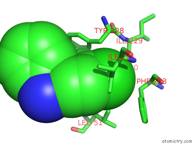
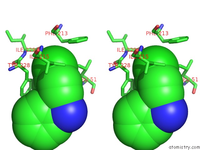
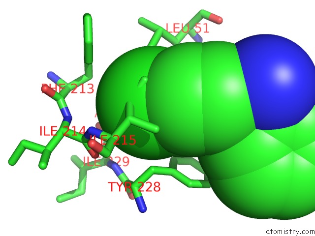
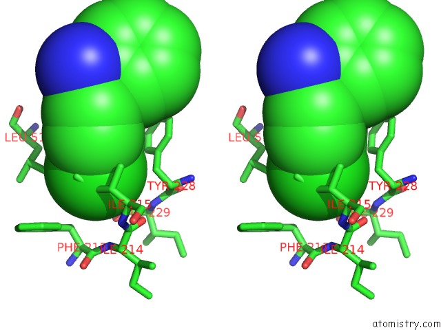
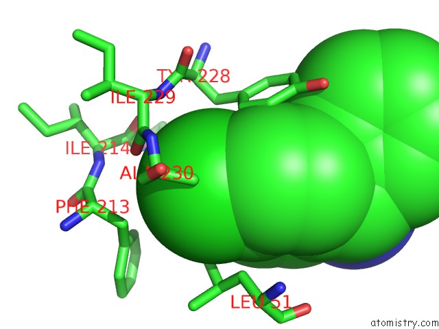
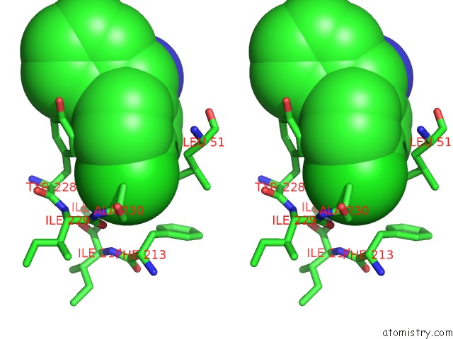
Chlorine binding site 1 out of 5 in 5wwv
Go back to
Chlorine binding site 1 out
of 5 in the Crystal Structure of Porcine Kidney D-Amino Acid Oxidase Mutant (I230A/R283G)
Mono view
Stereo pair view
Mono view
Stereo pair view
A full contact list of Chlorine with other atoms in the Cl binding
site number 1 of Crystal Structure of Porcine Kidney D-Amino Acid Oxidase Mutant (I230A/R283G) within 5.0Å range:
|
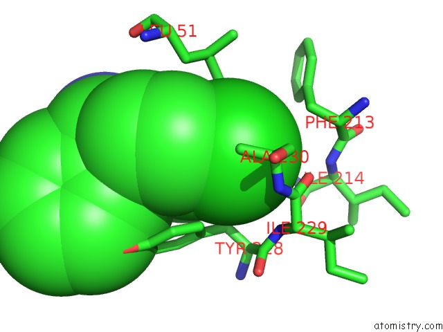
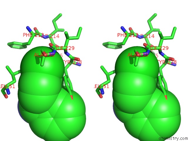
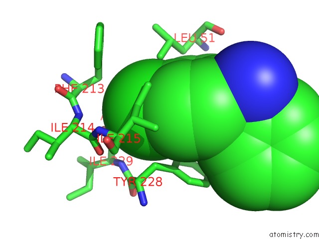
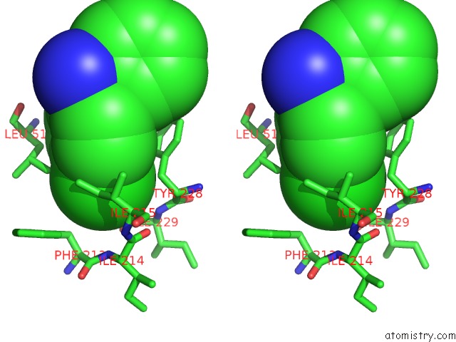
Chlorine binding site 2 out of 5 in 5wwv
Go back to
Chlorine binding site 2 out
of 5 in the Crystal Structure of Porcine Kidney D-Amino Acid Oxidase Mutant (I230A/R283G)
Mono view
Stereo pair view
Mono view
Stereo pair view
A full contact list of Chlorine with other atoms in the Cl binding
site number 2 of Crystal Structure of Porcine Kidney D-Amino Acid Oxidase Mutant (I230A/R283G) within 5.0Å range:
|
Chlorine binding site 3 out of 5 in 5wwv
Go back to
Chlorine binding site 3 out
of 5 in the Crystal Structure of Porcine Kidney D-Amino Acid Oxidase Mutant (I230A/R283G)
Mono view
Stereo pair view
Mono view
Stereo pair view
A full contact list of Chlorine with other atoms in the Cl binding
site number 3 of Crystal Structure of Porcine Kidney D-Amino Acid Oxidase Mutant (I230A/R283G) within 5.0Å range:
|
Chlorine binding site 4 out of 5 in 5wwv
Go back to
Chlorine binding site 4 out
of 5 in the Crystal Structure of Porcine Kidney D-Amino Acid Oxidase Mutant (I230A/R283G)
Mono view
Stereo pair view
Mono view
Stereo pair view
A full contact list of Chlorine with other atoms in the Cl binding
site number 4 of Crystal Structure of Porcine Kidney D-Amino Acid Oxidase Mutant (I230A/R283G) within 5.0Å range:
|
Chlorine binding site 5 out of 5 in 5wwv
Go back to
Chlorine binding site 5 out
of 5 in the Crystal Structure of Porcine Kidney D-Amino Acid Oxidase Mutant (I230A/R283G)
Mono view
Stereo pair view
Mono view
Stereo pair view
A full contact list of Chlorine with other atoms in the Cl binding
site number 5 of Crystal Structure of Porcine Kidney D-Amino Acid Oxidase Mutant (I230A/R283G) within 5.0Å range:
|
Reference:
K.Yasukawa,
F.Motojima,
A.Ohno,
Y.Asano.
Tailoring D-Amino Acid Oxidase From the Pid Kidney to R-Stereoselective Amine Oxidase and Its Use in the Deracemization of 4-Chlorobenzhydrylamine To Be Published.
Page generated: Fri Jul 26 20:43:15 2024
Last articles
Cl in 2WERCl in 2WD7
Cl in 2WEH
Cl in 2WDT
Cl in 2WD0
Cl in 2WD3
Cl in 2WCZ
Cl in 2WBK
Cl in 2WBF
Cl in 2WCG