Chlorine »
PDB 5wt2-5x2o »
5x23 »
Chlorine in PDB 5x23: Crystal Structure of CYP2C9 Genetic Variant A477T (*30) in Complex with Multiple Losartan Molecules
Enzymatic activity of Crystal Structure of CYP2C9 Genetic Variant A477T (*30) in Complex with Multiple Losartan Molecules
All present enzymatic activity of Crystal Structure of CYP2C9 Genetic Variant A477T (*30) in Complex with Multiple Losartan Molecules:
1.14.13.48; 1.14.13.49; 1.14.13.80; 1.14.99.38;
1.14.13.48; 1.14.13.49; 1.14.13.80; 1.14.99.38;
Protein crystallography data
The structure of Crystal Structure of CYP2C9 Genetic Variant A477T (*30) in Complex with Multiple Losartan Molecules, PDB code: 5x23
was solved by
K.Maekawa,
M.Adachi,
M.B.Shah,
with X-Ray Crystallography technique. A brief refinement statistics is given in the table below:
| Resolution Low / High (Å) | 45.51 / 2.00 |
| Space group | I 2 2 2 |
| Cell size a, b, c (Å), α, β, γ (°) | 74.952, 142.273, 161.831, 90.00, 90.00, 90.00 |
| R / Rfree (%) | 21.7 / 25.3 |
Other elements in 5x23:
The structure of Crystal Structure of CYP2C9 Genetic Variant A477T (*30) in Complex with Multiple Losartan Molecules also contains other interesting chemical elements:
| Potassium | (K) | 1 atom |
| Iron | (Fe) | 1 atom |
Chlorine Binding Sites:
The binding sites of Chlorine atom in the Crystal Structure of CYP2C9 Genetic Variant A477T (*30) in Complex with Multiple Losartan Molecules
(pdb code 5x23). This binding sites where shown within
5.0 Angstroms radius around Chlorine atom.
In total 4 binding sites of Chlorine where determined in the Crystal Structure of CYP2C9 Genetic Variant A477T (*30) in Complex with Multiple Losartan Molecules, PDB code: 5x23:
Jump to Chlorine binding site number: 1; 2; 3; 4;
In total 4 binding sites of Chlorine where determined in the Crystal Structure of CYP2C9 Genetic Variant A477T (*30) in Complex with Multiple Losartan Molecules, PDB code: 5x23:
Jump to Chlorine binding site number: 1; 2; 3; 4;
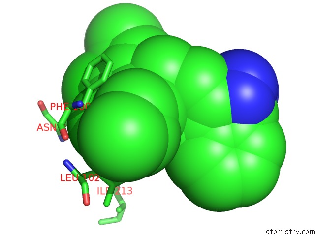
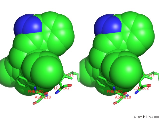
Chlorine binding site 1 out of 4 in 5x23
Go back to
Chlorine binding site 1 out
of 4 in the Crystal Structure of CYP2C9 Genetic Variant A477T (*30) in Complex with Multiple Losartan Molecules
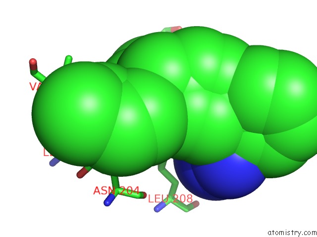
Mono view
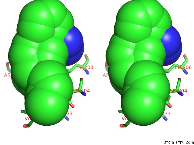
Stereo pair view
Mono view
Stereo pair view
A full contact list of Chlorine with other atoms in the Cl binding
site number 1 of Crystal Structure of CYP2C9 Genetic Variant A477T (*30) in Complex with Multiple Losartan Molecules within 5.0Å range:
|
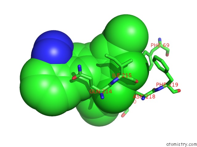
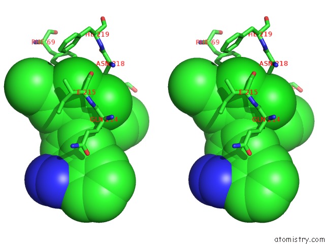
Chlorine binding site 2 out of 4 in 5x23
Go back to
Chlorine binding site 2 out
of 4 in the Crystal Structure of CYP2C9 Genetic Variant A477T (*30) in Complex with Multiple Losartan Molecules
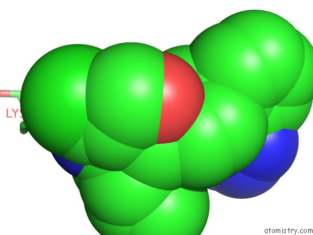
Mono view
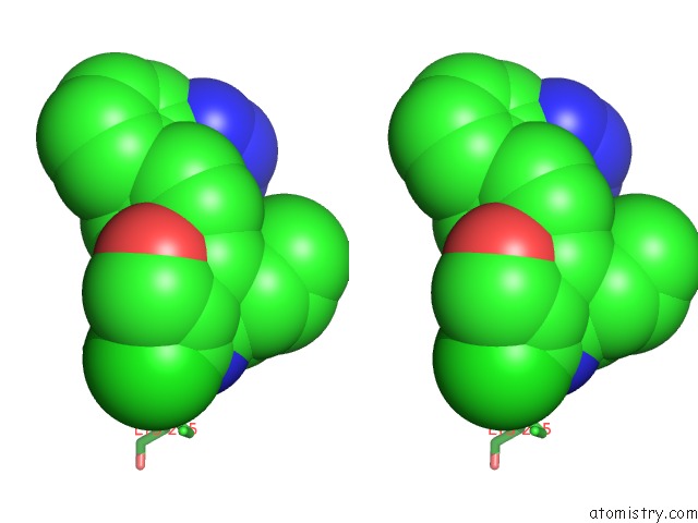
Stereo pair view
Mono view
Stereo pair view
A full contact list of Chlorine with other atoms in the Cl binding
site number 2 of Crystal Structure of CYP2C9 Genetic Variant A477T (*30) in Complex with Multiple Losartan Molecules within 5.0Å range:
|
Chlorine binding site 3 out of 4 in 5x23
Go back to
Chlorine binding site 3 out
of 4 in the Crystal Structure of CYP2C9 Genetic Variant A477T (*30) in Complex with Multiple Losartan Molecules
Mono view
Stereo pair view
Mono view
Stereo pair view
A full contact list of Chlorine with other atoms in the Cl binding
site number 3 of Crystal Structure of CYP2C9 Genetic Variant A477T (*30) in Complex with Multiple Losartan Molecules within 5.0Å range:
|
Chlorine binding site 4 out of 4 in 5x23
Go back to
Chlorine binding site 4 out
of 4 in the Crystal Structure of CYP2C9 Genetic Variant A477T (*30) in Complex with Multiple Losartan Molecules
Mono view
Stereo pair view
Mono view
Stereo pair view
A full contact list of Chlorine with other atoms in the Cl binding
site number 4 of Crystal Structure of CYP2C9 Genetic Variant A477T (*30) in Complex with Multiple Losartan Molecules within 5.0Å range:
|
Reference:
K.Maekawa,
M.Adachi,
Y.Matsuzawa,
Q.Zhang,
R.Kuroki,
Y.Saito,
M.B.Shah.
Structural Basis of Single-Nucleotide Polymorphisms in Cytochrome P450 2C9 Biochemistry V. 56 5476 2017.
ISSN: ISSN 1520-4995
PubMed: 28972767
DOI: 10.1021/ACS.BIOCHEM.7B00795
Page generated: Fri Jul 26 20:45:46 2024
ISSN: ISSN 1520-4995
PubMed: 28972767
DOI: 10.1021/ACS.BIOCHEM.7B00795
Last articles
Zn in 9MJ5Zn in 9HNW
Zn in 9G0L
Zn in 9FNE
Zn in 9DZN
Zn in 9E0I
Zn in 9D32
Zn in 9DAK
Zn in 8ZXC
Zn in 8ZUF