Chlorine »
PDB 5x2o-5xif »
5xev »
Chlorine in PDB 5xev: Crystal Structure of A Novel Xaa-Pro Dipeptidase From Deinococcus Radiodurans
Enzymatic activity of Crystal Structure of A Novel Xaa-Pro Dipeptidase From Deinococcus Radiodurans
All present enzymatic activity of Crystal Structure of A Novel Xaa-Pro Dipeptidase From Deinococcus Radiodurans:
3.4.13.9;
3.4.13.9;
Protein crystallography data
The structure of Crystal Structure of A Novel Xaa-Pro Dipeptidase From Deinococcus Radiodurans, PDB code: 5xev
was solved by
V.N.Are,
A.Kumar,
B.Ghosh,
R.D.Makde,
with X-Ray Crystallography technique. A brief refinement statistics is given in the table below:
| Resolution Low / High (Å) | 31.96 / 1.40 |
| Space group | C 1 2 1 |
| Cell size a, b, c (Å), α, β, γ (°) | 90.670, 91.879, 53.849, 90.00, 98.78, 90.00 |
| R / Rfree (%) | 14.6 / 16.4 |
Other elements in 5xev:
The structure of Crystal Structure of A Novel Xaa-Pro Dipeptidase From Deinococcus Radiodurans also contains other interesting chemical elements:
| Zinc | (Zn) | 2 atoms |
Chlorine Binding Sites:
The binding sites of Chlorine atom in the Crystal Structure of A Novel Xaa-Pro Dipeptidase From Deinococcus Radiodurans
(pdb code 5xev). This binding sites where shown within
5.0 Angstroms radius around Chlorine atom.
In total only one binding site of Chlorine was determined in the Crystal Structure of A Novel Xaa-Pro Dipeptidase From Deinococcus Radiodurans, PDB code: 5xev:
In total only one binding site of Chlorine was determined in the Crystal Structure of A Novel Xaa-Pro Dipeptidase From Deinococcus Radiodurans, PDB code: 5xev:
Chlorine binding site 1 out of 1 in 5xev
Go back to
Chlorine binding site 1 out
of 1 in the Crystal Structure of A Novel Xaa-Pro Dipeptidase From Deinococcus Radiodurans
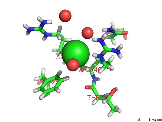
Mono view
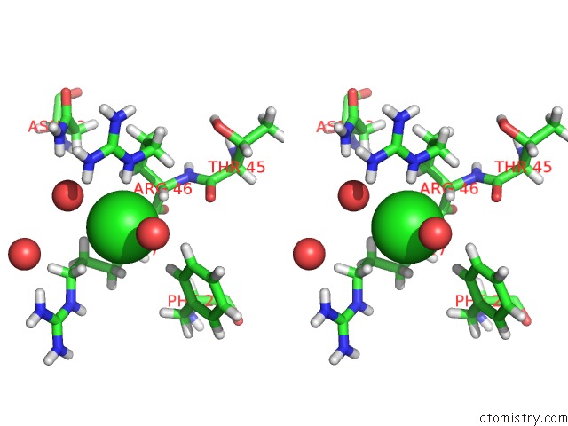
Stereo pair view
Mono view
Stereo pair view
A full contact list of Chlorine with other atoms in the Cl binding
site number 1 of Crystal Structure of A Novel Xaa-Pro Dipeptidase From Deinococcus Radiodurans within 5.0Å range:
|
Reference:
V.N.Are,
S.N.Jamdar,
B.Ghosh,
V.D.Goyal,
A.Kumar,
S.Neema,
R.Gadre,
R.D.Makde.
Crystal Structure of A Novel Prolidase From Deinococcus Radiodurans Identifies New Subfamily of Bacterial Prolidases. Proteins V. 85 2239 2017.
ISSN: ESSN 1097-0134
PubMed: 28929533
DOI: 10.1002/PROT.25389
Page generated: Fri Jul 26 20:54:18 2024
ISSN: ESSN 1097-0134
PubMed: 28929533
DOI: 10.1002/PROT.25389
Last articles
Zn in 9JYWZn in 9IR4
Zn in 9IR3
Zn in 9GMX
Zn in 9GMW
Zn in 9JEJ
Zn in 9ERF
Zn in 9ERE
Zn in 9EGV
Zn in 9EGW