Chlorine »
PDB 5x2p-5xih »
5xh6 »
Chlorine in PDB 5xh6: Crystal Structure of the Acidaminococcus Sp. BV3L6 CPF1 Rvr Variant in Complex with Crrna and Target Dna (Tata Pam)
Protein crystallography data
The structure of Crystal Structure of the Acidaminococcus Sp. BV3L6 CPF1 Rvr Variant in Complex with Crrna and Target Dna (Tata Pam), PDB code: 5xh6
was solved by
H.Nishimasu,
T.Yamano,
R.Ishitani,
O.Nureki,
with X-Ray Crystallography technique. A brief refinement statistics is given in the table below:
| Resolution Low / High (Å) | 39.99 / 2.00 |
| Space group | P 21 21 21 |
| Cell size a, b, c (Å), α, β, γ (°) | 81.162, 133.676, 199.633, 90.00, 90.00, 90.00 |
| R / Rfree (%) | 17.5 / 21 |
Other elements in 5xh6:
The structure of Crystal Structure of the Acidaminococcus Sp. BV3L6 CPF1 Rvr Variant in Complex with Crrna and Target Dna (Tata Pam) also contains other interesting chemical elements:
| Sodium | (Na) | 5 atoms |
Chlorine Binding Sites:
The binding sites of Chlorine atom in the Crystal Structure of the Acidaminococcus Sp. BV3L6 CPF1 Rvr Variant in Complex with Crrna and Target Dna (Tata Pam)
(pdb code 5xh6). This binding sites where shown within
5.0 Angstroms radius around Chlorine atom.
In total only one binding site of Chlorine was determined in the Crystal Structure of the Acidaminococcus Sp. BV3L6 CPF1 Rvr Variant in Complex with Crrna and Target Dna (Tata Pam), PDB code: 5xh6:
In total only one binding site of Chlorine was determined in the Crystal Structure of the Acidaminococcus Sp. BV3L6 CPF1 Rvr Variant in Complex with Crrna and Target Dna (Tata Pam), PDB code: 5xh6:
Chlorine binding site 1 out of 1 in 5xh6
Go back to
Chlorine binding site 1 out
of 1 in the Crystal Structure of the Acidaminococcus Sp. BV3L6 CPF1 Rvr Variant in Complex with Crrna and Target Dna (Tata Pam)
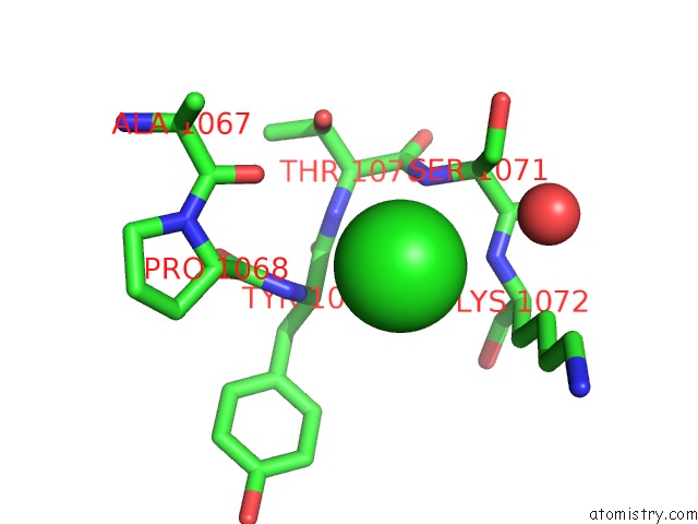
Mono view
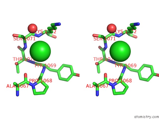
Stereo pair view
Mono view
Stereo pair view
A full contact list of Chlorine with other atoms in the Cl binding
site number 1 of Crystal Structure of the Acidaminococcus Sp. BV3L6 CPF1 Rvr Variant in Complex with Crrna and Target Dna (Tata Pam) within 5.0Å range:
|
Reference:
H.Nishimasu,
T.Yamano,
L.Gao,
F.Zhang,
R.Ishitani,
O.Nureki.
Structural Basis For the Altered Pam Recognition By Engineered Crispr-CPF1 Mol. Cell V. 67 139 2017.
ISSN: ISSN 1097-4164
PubMed: 28595896
DOI: 10.1016/J.MOLCEL.2017.04.019
Page generated: Fri Jul 26 20:55:59 2024
ISSN: ISSN 1097-4164
PubMed: 28595896
DOI: 10.1016/J.MOLCEL.2017.04.019
Last articles
Zn in 9J0NZn in 9J0O
Zn in 9J0P
Zn in 9FJX
Zn in 9EKB
Zn in 9C0F
Zn in 9CAH
Zn in 9CH0
Zn in 9CH3
Zn in 9CH1