Atomistry » Chlorine » PDB 5xii-5xvf » 5xpe
Atomistry »
  Chlorine »
    PDB 5xii-5xvf »
      5xpe »

Chlorine in PDB 5xpe: Neutron Structure of the T26H Mutant of T4 Lysozyme

Enzymatic activity of Neutron Structure of the T26H Mutant of T4 Lysozyme

All present enzymatic activity of Neutron Structure of the T26H Mutant of T4 Lysozyme:
3.2.1.17;

Protein crystallography data

The structure of Neutron Structure of the T26H Mutant of T4 Lysozyme, PDB code: 5xpe was solved by T.Hiromoto, R.Kuroki, with X-Ray Crystallography technique. A brief refinement statistics is given in the table below:

Resolution Low / High (Å) N/A / 1.65
Space group P 32 2 1
Cell size a, b, c (Å), α, β, γ (°) 61.230, 61.230, 96.791, 90.00, 90.00, 120.00
R / Rfree (%) 22.5 / 27.8

Other elements in 5xpe:

The structure of Neutron Structure of the T26H Mutant of T4 Lysozyme also contains other interesting chemical elements:

Sodium (Na) 1 atom

Chlorine Binding Sites:

The binding sites of Chlorine atom in the Neutron Structure of the T26H Mutant of T4 Lysozyme (pdb code 5xpe). This binding sites where shown within 5.0 Angstroms radius around Chlorine atom.
In total 4 binding sites of Chlorine where determined in the Neutron Structure of the T26H Mutant of T4 Lysozyme, PDB code: 5xpe:
Jump to Chlorine binding site number: 1; 2; 3; 4;

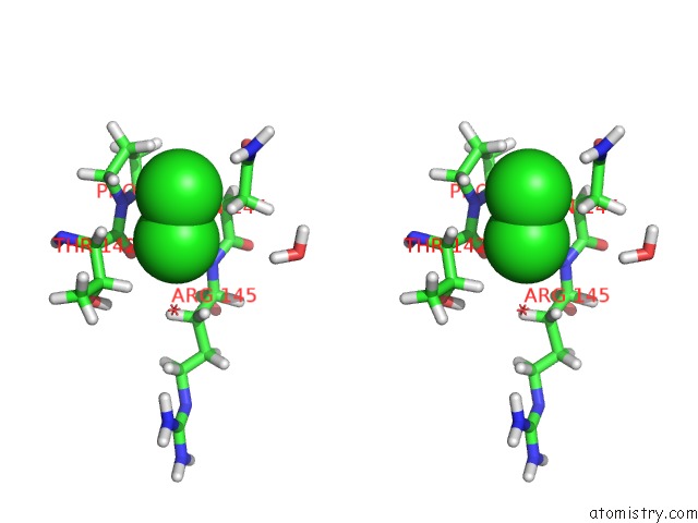
Chlorine binding site 1 out of 4 in 5xpe

Go back to Chlorine Binding Sites List in 5xpe
Chlorine binding site 1 out of 4 in the Neutron Structure of the T26H Mutant of T4 Lysozyme


Mono view


Stereo pair view

A full contact list of Chlorine with other atoms in the Cl binding site number 1 of Neutron Structure of the T26H Mutant of T4 Lysozyme within 5.0Å range:
probe atom residue distance (Å) B Occ
A:Cl202

b:35.6
occ:0.67
CL A:CL202 0.0 35.6 0.7
CL A:CL202 2.0 40.1 0.3
D A:ARG145 2.4 33.8 1.0
D A:ASN144 2.5 28.2 1.0
DB2 A:ASN144 2.7 27.2 1.0
DB A:THR142 2.9 27.2 1.0
DA A:THR142 3.1 24.5 1.0
O A:DOD386 3.1 46.3 1.0
N A:ASN144 3.3 26.2 1.0
N A:ARG145 3.3 28.2 1.0
C A:THR142 3.4 28.9 1.0
DB2 A:ARG145 3.5 34.4 1.0
CA A:THR142 3.6 25.9 1.0
DD2 A:PRO143 3.6 30.8 1.0
CB A:ASN144 3.7 29.8 1.0
O A:THR142 3.7 28.4 1.0
CB A:THR142 3.7 31.2 1.0
N A:PRO143 3.7 26.1 1.0
CA A:ASN144 3.8 27.9 1.0
DB3 A:ARG145 4.0 20.2 1.0
C A:ASN144 4.1 28.8 1.0
CB A:ARG145 4.1 26.2 1.0
DD21 A:ASN144 4.1 43.9 1.0
D2 A:DOD378 4.1 53.2 1.0
DG23 A:THR142 4.1 30.0 1.0
C A:PRO143 4.2 25.5 1.0
CD A:PRO143 4.2 29.9 1.0
CA A:ARG145 4.3 27.1 1.0
DG2 A:PRO143 4.3 26.9 1.0
DB3 A:ASN144 4.4 25.5 1.0
CG2 A:THR142 4.4 30.6 1.0
CA A:PRO143 4.5 26.6 1.0
D A:ALA146 4.6 35.6 1.0
CG A:ASN144 4.6 37.9 1.0
DG22 A:THR142 4.6 38.3 1.0
ND2 A:ASN144 4.8 43.3 1.0
O A:DOD316 4.8 45.1 1.0
CG A:PRO143 4.8 28.1 1.0
DG1 A:THR142 4.8 34.9 1.0
DA A:ASN144 4.9 45.0 1.0
OG1 A:THR142 4.9 31.4 1.0
N A:THR142 5.0 27.4 1.0
O A:DOD378 5.0 60.6 1.0
DA A:ARG145 5.0 23.7 1.0

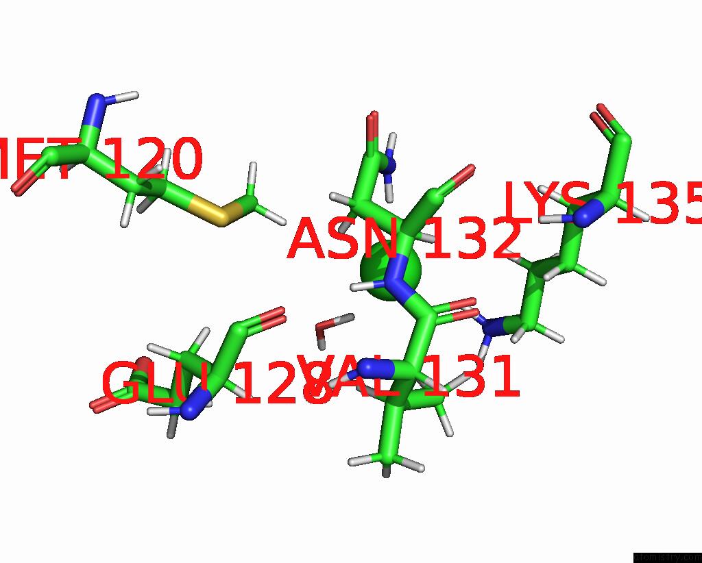
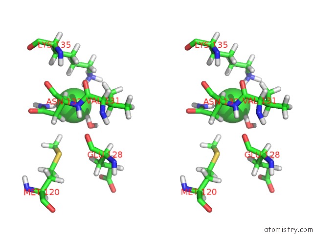
Chlorine binding site 2 out of 4 in 5xpe

Go back to Chlorine Binding Sites List in 5xpe
Chlorine binding site 2 out of 4 in the Neutron Structure of the T26H Mutant of T4 Lysozyme


Mono view


Stereo pair view

A full contact list of Chlorine with other atoms in the Cl binding site number 2 of Neutron Structure of the T26H Mutant of T4 Lysozyme within 5.0Å range:
probe atom residue distance (Å) B Occ
A:Cl202

b:40.1
occ:0.33
CL A:CL202 0.0 40.1 0.3
CL A:CL202 2.0 35.6 0.7
D A:ASN144 2.3 28.2 1.0
DB2 A:ASN144 2.6 27.2 1.0
DD21 A:ASN144 2.7 43.9 1.0
DD2 A:PRO143 2.9 30.8 1.0
DG2 A:PRO143 3.1 26.9 1.0
N A:ASN144 3.3 26.2 1.0
CB A:ASN144 3.5 29.8 1.0
ND2 A:ASN144 3.5 43.3 1.0
DA A:THR142 3.6 24.5 1.0
CD A:PRO143 3.7 29.9 1.0
CG A:ASN144 3.7 37.9 1.0
N A:PRO143 3.8 26.1 1.0
D A:ARG145 3.8 33.8 1.0
CG A:PRO143 3.9 28.1 1.0
CA A:ASN144 4.0 27.9 1.0
C A:THR142 4.2 28.9 1.0
C A:PRO143 4.3 25.5 1.0
DB3 A:ASN144 4.4 25.5 1.0
DD22 A:ASN144 4.4 47.4 1.0
CA A:THR142 4.4 25.9 1.0
CA A:PRO143 4.5 26.6 1.0
OD1 A:ASN144 4.6 45.2 1.0
DB A:THR142 4.6 27.2 1.0
N A:ARG145 4.6 28.2 1.0
DG3 A:PRO143 4.6 27.9 1.0
DD3 A:PRO143 4.6 28.7 1.0
D2 A:DOD378 4.7 53.2 1.0
CB A:PRO143 4.7 31.8 1.0
O A:DOD386 4.8 46.3 1.0
DA A:ASN144 4.8 45.0 1.0
O A:THR142 4.8 28.4 1.0
C A:ASN144 4.9 28.8 1.0
DB2 A:PRO143 4.9 29.2 1.0

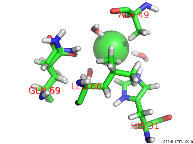
Chlorine binding site 3 out of 4 in 5xpe

Go back to Chlorine Binding Sites List in 5xpe
Chlorine binding site 3 out of 4 in the Neutron Structure of the T26H Mutant of T4 Lysozyme


Mono view


Stereo pair view

A full contact list of Chlorine with other atoms in the Cl binding site number 3 of Neutron Structure of the T26H Mutant of T4 Lysozyme within 5.0Å range:
probe atom residue distance (Å) B Occ
A:Cl203

b:55.4
occ:0.76
DD22 A:ASN132 2.1 38.6 1.0
D2 A:DOD348 2.1 50.8 1.0
DZ1 A:LYS135 2.4 56.2 1.0
O A:DOD348 2.8 48.9 1.0
DB2 A:ASN132 2.9 37.5 1.0
D1 A:DOD348 2.9 54.3 1.0
DA A:ASN132 3.1 45.4 1.0
ND2 A:ASN132 3.1 38.0 1.0
DD2 A:LYS135 3.3 54.4 1.0
NZ A:LYS135 3.4 74.5 1.0
DE1 A:MET120 3.4 44.4 1.0
DG12 A:VAL131 3.5 37.6 1.0
CB A:ASN132 3.6 29.2 1.0
DZ3 A:LYS135 3.7 59.1 1.0
CA A:ASN132 3.7 30.4 1.0
DD21 A:ASN132 3.8 38.5 1.0
CG A:ASN132 3.9 32.3 1.0
DZ2 A:LYS135 4.0 68.1 1.0
N A:ASN132 4.2 30.8 1.0
CE A:LYS135 4.2 72.0 1.0
CD A:LYS135 4.2 47.0 1.0
DE3 A:LYS135 4.2 59.5 1.0
D A:ASN132 4.4 33.1 1.0
CG1 A:VAL131 4.5 44.2 1.0
CE A:MET120 4.5 42.8 1.0
DB3 A:ASN132 4.6 26.9 1.0
DG11 A:VAL131 4.6 50.7 1.0
DB A:VAL131 4.6 34.5 1.0
C A:VAL131 4.7 34.7 1.0
DD3 A:LYS135 4.8 38.2 1.0
O A:GLU128 4.9 33.3 1.0
O A:VAL131 4.9 35.0 1.0
DE3 A:MET120 4.9 48.0 1.0
DE2 A:MET120 5.0 48.7 1.0

Chlorine binding site 4 out of 4 in 5xpe

Go back to Chlorine Binding Sites List in 5xpe
Chlorine binding site 4 out of 4 in the Neutron Structure of the T26H Mutant of T4 Lysozyme


Mono view


Stereo pair view

A full contact list of Chlorine with other atoms in the Cl binding site number 4 of Neutron Structure of the T26H Mutant of T4 Lysozyme within 5.0Å range:
probe atom residue distance (Å) B Occ
A:Cl204

b:47.3
occ:0.58
D2 A:DOD364 2.3 28.2 1.0
D1 A:DOD322 2.3 35.2 1.0
D1 A:DOD312 3.0 57.6 1.0
DB3 A:ALA49 3.0 38.2 1.0
DE1 A:HIS31 3.2 46.6 1.0
O A:DOD364 3.2 35.6 1.0
O A:DOD322 3.2 44.5 1.0
O A:DOD312 3.3 37.8 1.0
DE2 A:HIS31 3.5 40.4 1.0
D1 A:DOD364 3.8 28.5 1.0
DA A:ALA49 3.8 27.1 1.0
D2 A:DOD322 3.8 37.7 1.0
CE1 A:HIS31 3.8 33.1 1.0
CB A:ALA49 3.9 30.4 1.0
DD23 A:LEU66 4.0 33.5 1.0
DE22 A:GLN69 4.0 30.5 0.5
NE2 A:HIS31 4.0 31.1 1.0
DB1 A:ALA49 4.1 36.9 1.0
D2 A:DOD312 4.2 43.5 1.0
OE1 A:GLN69 4.4 33.7 0.5
DD22 A:LEU66 4.4 37.3 1.0
CA A:ALA49 4.5 30.1 1.0
CD2 A:LEU66 4.6 34.5 1.0
DE21 A:GLN69 4.7 29.9 0.5
NE2 A:GLN69 4.7 32.0 0.5
DB2 A:ALA49 4.8 39.0 1.0
DD21 A:LEU66 5.0 29.1 1.0

Reference:

T.Hiromoto, F.Meilleur, R.Shimizu, C.Shibazaki, M.Adachi, T.Tamada, R.Kuroki. Neutron Structure of the T26H Mutant of T4 Phage Lysozyme Provides Insight Into the Catalytic Activity of the Mutant Enzyme and How It Differs From That of Wild Type. Protein Sci. V. 26 1953 2017.
ISSN: ESSN 1469-896X
PubMed: 28707339
DOI: 10.1002/PRO.3230
Page generated: Sat Jul 12 10:44:03 2025

Last articles

Fe in 2YXO
Fe in 2YRS
Fe in 2YXC
Fe in 2YNM
Fe in 2YVJ
Fe in 2YP1
Fe in 2YU2
Fe in 2YU1
Fe in 2YQB
Fe in 2YOO
© Copyright 2008-2020 by atomistry.com
Home   |    Site Map   |    Copyright   |    Contact us   |    Privacy