Chlorine »
PDB 5xvg-5y7d »
5xyr »
Chlorine in PDB 5xyr: Crystal Structure of A Serine Protease From Streptococcus Species
Protein crystallography data
The structure of Crystal Structure of A Serine Protease From Streptococcus Species, PDB code: 5xyr
was solved by
C.Jobichen,
J.Sivaraman,
with X-Ray Crystallography technique. A brief refinement statistics is given in the table below:
| Resolution Low / High (Å) | 19.99 / 2.80 |
| Space group | P 62 2 2 |
| Cell size a, b, c (Å), α, β, γ (°) | 191.625, 191.625, 250.956, 90.00, 90.00, 120.00 |
| R / Rfree (%) | 20.9 / 25.7 |
Other elements in 5xyr:
The structure of Crystal Structure of A Serine Protease From Streptococcus Species also contains other interesting chemical elements:
| Calcium | (Ca) | 3 atoms |
Chlorine Binding Sites:
The binding sites of Chlorine atom in the Crystal Structure of A Serine Protease From Streptococcus Species
(pdb code 5xyr). This binding sites where shown within
5.0 Angstroms radius around Chlorine atom.
In total only one binding site of Chlorine was determined in the Crystal Structure of A Serine Protease From Streptococcus Species, PDB code: 5xyr:
In total only one binding site of Chlorine was determined in the Crystal Structure of A Serine Protease From Streptococcus Species, PDB code: 5xyr:
Chlorine binding site 1 out of 1 in 5xyr
Go back to
Chlorine binding site 1 out
of 1 in the Crystal Structure of A Serine Protease From Streptococcus Species
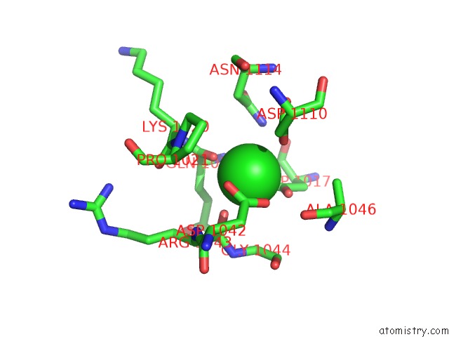
Mono view
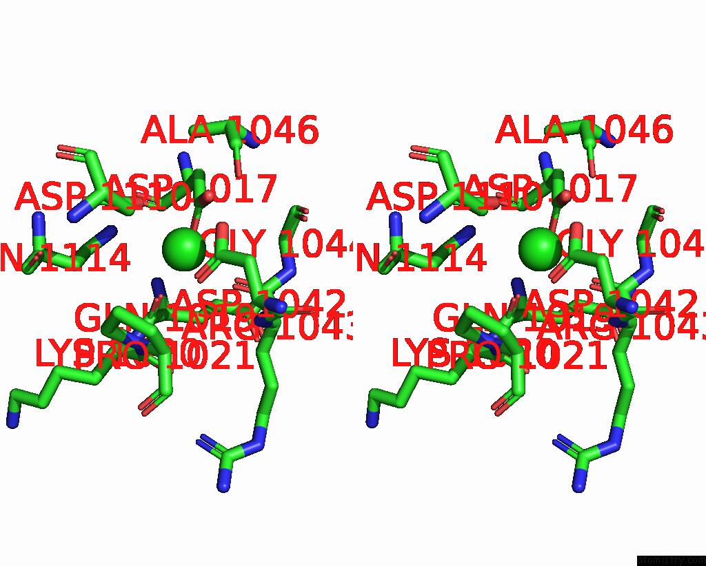
Stereo pair view
Mono view
Stereo pair view
A full contact list of Chlorine with other atoms in the Cl binding
site number 1 of Crystal Structure of A Serine Protease From Streptococcus Species within 5.0Å range:
|
Reference:
C.Jobichen,
Y.C.Tan,
M.T.Prabhakar,
D.Nayak,
D.Biswas,
N.S.Pannu,
E.Hanski,
J.Sivaraman.
Structure of Scpc, A Virulence Protease Fromstreptococcus Pyogenes, Reveals the Functional Domains and Maturation Mechanism. Biochem. J. V. 475 2847 2018.
ISSN: ESSN 1470-8728
PubMed: 30049896
DOI: 10.1042/BCJ20180145
Page generated: Fri Jul 26 21:12:44 2024
ISSN: ESSN 1470-8728
PubMed: 30049896
DOI: 10.1042/BCJ20180145
Last articles
Zn in 9J0NZn in 9J0O
Zn in 9J0P
Zn in 9FJX
Zn in 9EKB
Zn in 9C0F
Zn in 9CAH
Zn in 9CH0
Zn in 9CH3
Zn in 9CH1