Chlorine »
PDB 5xvg-5y7d »
5y0m »
Chlorine in PDB 5y0m: Structure of 6-Aminohexanoate-Oligomer Hydrolase From Arthrobacter Sp. KI72., D36A/D122G/H130Y/E263Q Muntant
Protein crystallography data
The structure of Structure of 6-Aminohexanoate-Oligomer Hydrolase From Arthrobacter Sp. KI72., D36A/D122G/H130Y/E263Q Muntant, PDB code: 5y0m
was solved by
S.Negoro,
N.Shibata,
K.Nagai,
Y.Higuchi,
with X-Ray Crystallography technique. A brief refinement statistics is given in the table below:
| Resolution Low / High (Å) | 72.21 / 1.03 |
| Space group | C 2 2 21 |
| Cell size a, b, c (Å), α, β, γ (°) | 70.418, 144.424, 128.268, 90.00, 90.00, 90.00 |
| R / Rfree (%) | 11.6 / 13.4 |
Chlorine Binding Sites:
The binding sites of Chlorine atom in the Structure of 6-Aminohexanoate-Oligomer Hydrolase From Arthrobacter Sp. KI72., D36A/D122G/H130Y/E263Q Muntant
(pdb code 5y0m). This binding sites where shown within
5.0 Angstroms radius around Chlorine atom.
In total 4 binding sites of Chlorine where determined in the Structure of 6-Aminohexanoate-Oligomer Hydrolase From Arthrobacter Sp. KI72., D36A/D122G/H130Y/E263Q Muntant, PDB code: 5y0m:
Jump to Chlorine binding site number: 1; 2; 3; 4;
In total 4 binding sites of Chlorine where determined in the Structure of 6-Aminohexanoate-Oligomer Hydrolase From Arthrobacter Sp. KI72., D36A/D122G/H130Y/E263Q Muntant, PDB code: 5y0m:
Jump to Chlorine binding site number: 1; 2; 3; 4;
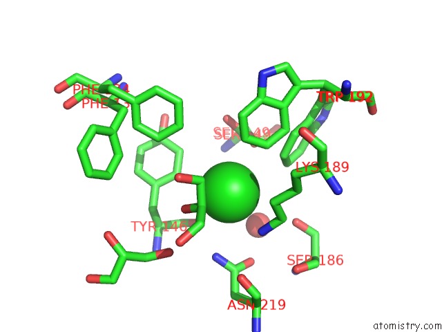
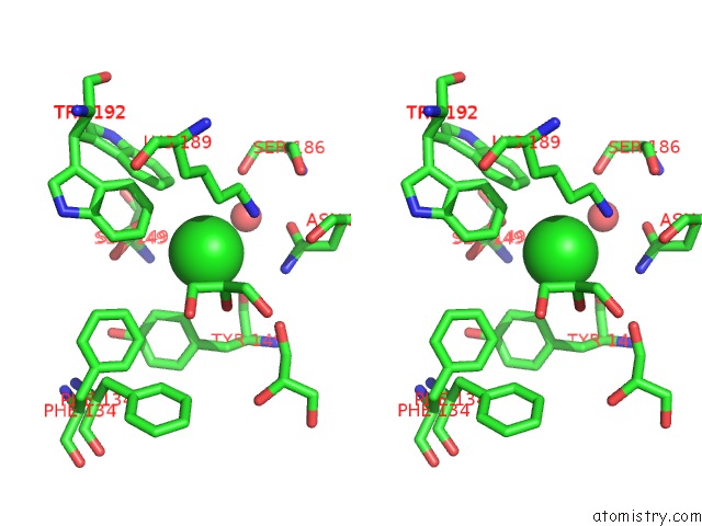
Chlorine binding site 1 out of 4 in 5y0m
Go back to
Chlorine binding site 1 out
of 4 in the Structure of 6-Aminohexanoate-Oligomer Hydrolase From Arthrobacter Sp. KI72., D36A/D122G/H130Y/E263Q Muntant
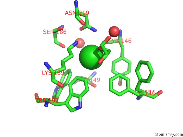
Mono view
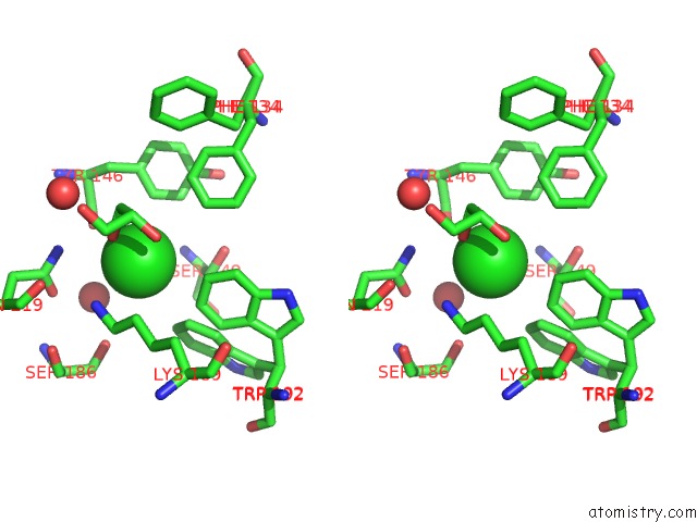
Stereo pair view
Mono view
Stereo pair view
A full contact list of Chlorine with other atoms in the Cl binding
site number 1 of Structure of 6-Aminohexanoate-Oligomer Hydrolase From Arthrobacter Sp. KI72., D36A/D122G/H130Y/E263Q Muntant within 5.0Å range:
|
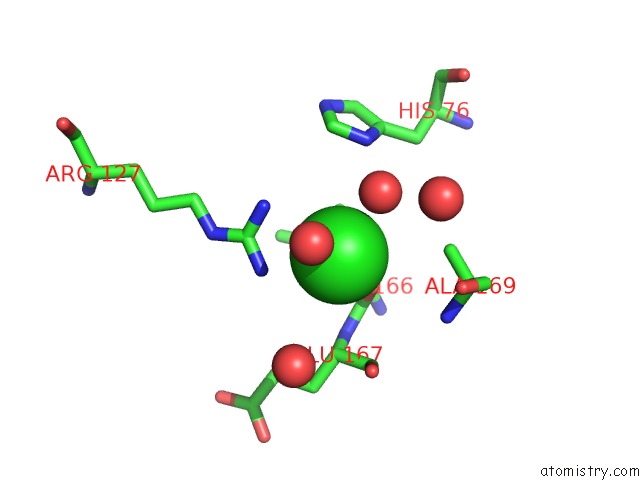
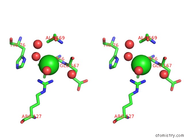
Chlorine binding site 2 out of 4 in 5y0m
Go back to
Chlorine binding site 2 out
of 4 in the Structure of 6-Aminohexanoate-Oligomer Hydrolase From Arthrobacter Sp. KI72., D36A/D122G/H130Y/E263Q Muntant
Mono view
Stereo pair view
Mono view
Stereo pair view
A full contact list of Chlorine with other atoms in the Cl binding
site number 2 of Structure of 6-Aminohexanoate-Oligomer Hydrolase From Arthrobacter Sp. KI72., D36A/D122G/H130Y/E263Q Muntant within 5.0Å range:
|
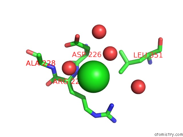
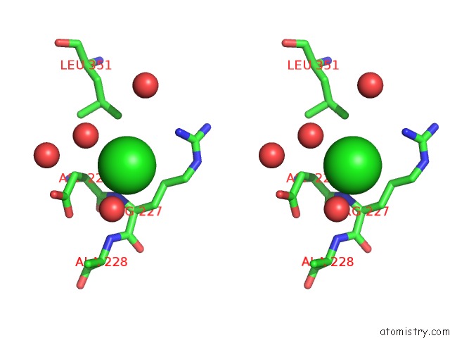
Chlorine binding site 3 out of 4 in 5y0m
Go back to
Chlorine binding site 3 out
of 4 in the Structure of 6-Aminohexanoate-Oligomer Hydrolase From Arthrobacter Sp. KI72., D36A/D122G/H130Y/E263Q Muntant
Mono view
Stereo pair view
Mono view
Stereo pair view
A full contact list of Chlorine with other atoms in the Cl binding
site number 3 of Structure of 6-Aminohexanoate-Oligomer Hydrolase From Arthrobacter Sp. KI72., D36A/D122G/H130Y/E263Q Muntant within 5.0Å range:
|
Chlorine binding site 4 out of 4 in 5y0m
Go back to
Chlorine binding site 4 out
of 4 in the Structure of 6-Aminohexanoate-Oligomer Hydrolase From Arthrobacter Sp. KI72., D36A/D122G/H130Y/E263Q Muntant
Mono view
Stereo pair view
Mono view
Stereo pair view
A full contact list of Chlorine with other atoms in the Cl binding
site number 4 of Structure of 6-Aminohexanoate-Oligomer Hydrolase From Arthrobacter Sp. KI72., D36A/D122G/H130Y/E263Q Muntant within 5.0Å range:
|
Reference:
S.Negoro,
N.Shibata,
Y.H.Lee,
I.Takehara,
R.Kinugasa,
K.Nagai,
Y.Tanaka,
D.I.Kato,
M.Takeo,
Y.Goto,
Y.Higuchi.
Structural Basis of the Correct Subunit Assembly, Aggregation, and Intracellular Degradation of Nylon Hydrolase Sci Rep V. 8 9725 2018.
ISSN: ESSN 2045-2322
PubMed: 29950566
DOI: 10.1038/S41598-018-27860-W
Page generated: Sat Jul 12 10:49:42 2025
ISSN: ESSN 2045-2322
PubMed: 29950566
DOI: 10.1038/S41598-018-27860-W
Last articles
F in 4J8WF in 4J8M
F in 4J6A
F in 4J6I
F in 4J53
F in 4J1K
F in 4J1I
F in 4J52
F in 4J1H
F in 4J1E