Chlorine »
PDB 5xvg-5y7d »
5y5e »
Chlorine in PDB 5y5e: Crystal Structure of Phospholipase A2 with Inhibitor
Enzymatic activity of Crystal Structure of Phospholipase A2 with Inhibitor
All present enzymatic activity of Crystal Structure of Phospholipase A2 with Inhibitor:
3.1.1.4;
3.1.1.4;
Protein crystallography data
The structure of Crystal Structure of Phospholipase A2 with Inhibitor, PDB code: 5y5e
was solved by
S.Hou,
J.Xu,
T.Xu,
J.Liu,
with X-Ray Crystallography technique. A brief refinement statistics is given in the table below:
| Resolution Low / High (Å) | 18.78 / 1.80 |
| Space group | P 21 2 2 |
| Cell size a, b, c (Å), α, β, γ (°) | 48.726, 61.063, 63.356, 90.00, 90.00, 90.00 |
| R / Rfree (%) | 19.3 / 22.5 |
Other elements in 5y5e:
The structure of Crystal Structure of Phospholipase A2 with Inhibitor also contains other interesting chemical elements:
| Fluorine | (F) | 3 atoms |
| Calcium | (Ca) | 1 atom |
| Sodium | (Na) | 1 atom |
Chlorine Binding Sites:
The binding sites of Chlorine atom in the Crystal Structure of Phospholipase A2 with Inhibitor
(pdb code 5y5e). This binding sites where shown within
5.0 Angstroms radius around Chlorine atom.
In total 5 binding sites of Chlorine where determined in the Crystal Structure of Phospholipase A2 with Inhibitor, PDB code: 5y5e:
Jump to Chlorine binding site number: 1; 2; 3; 4; 5;
In total 5 binding sites of Chlorine where determined in the Crystal Structure of Phospholipase A2 with Inhibitor, PDB code: 5y5e:
Jump to Chlorine binding site number: 1; 2; 3; 4; 5;
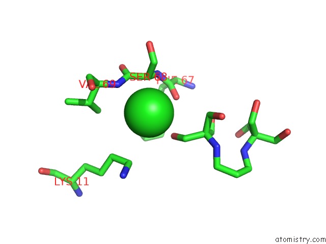
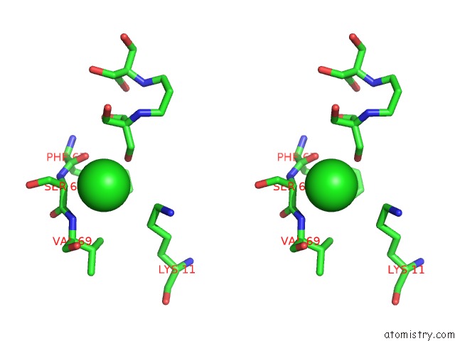
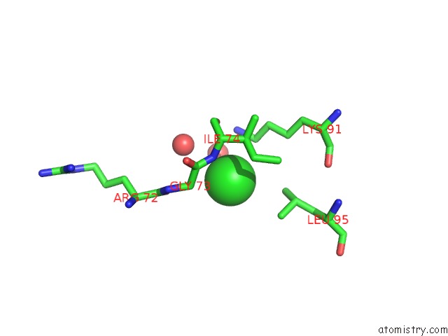
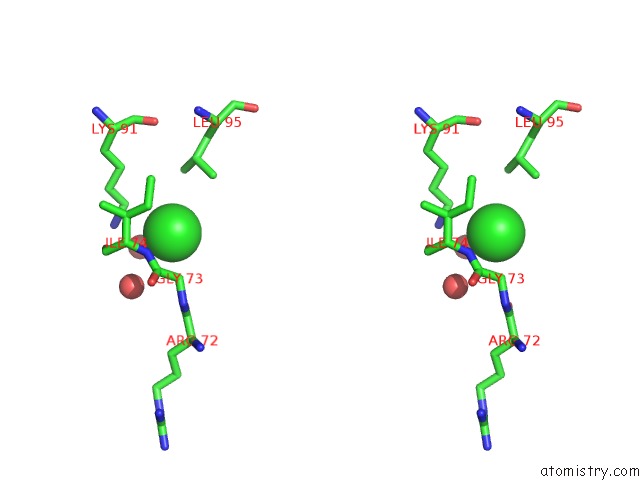
Chlorine binding site 1 out of 5 in 5y5e
Go back to
Chlorine binding site 1 out
of 5 in the Crystal Structure of Phospholipase A2 with Inhibitor
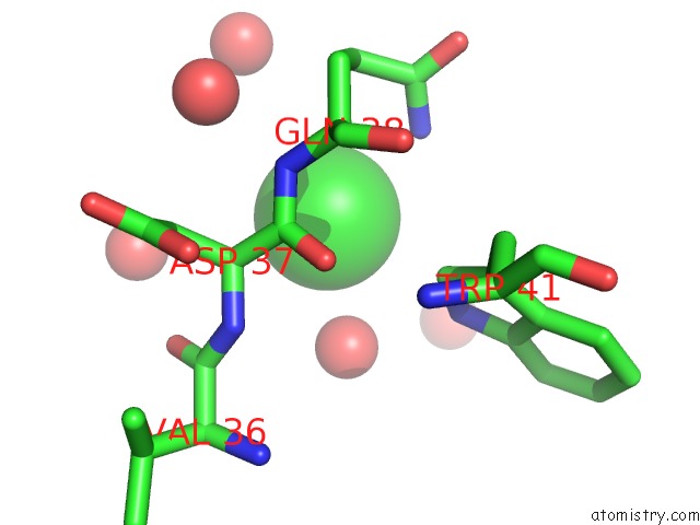
Mono view
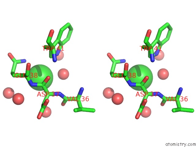
Stereo pair view
Mono view
Stereo pair view
A full contact list of Chlorine with other atoms in the Cl binding
site number 1 of Crystal Structure of Phospholipase A2 with Inhibitor within 5.0Å range:
|
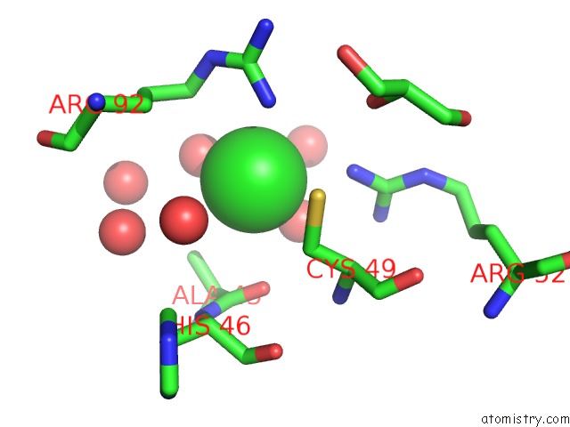
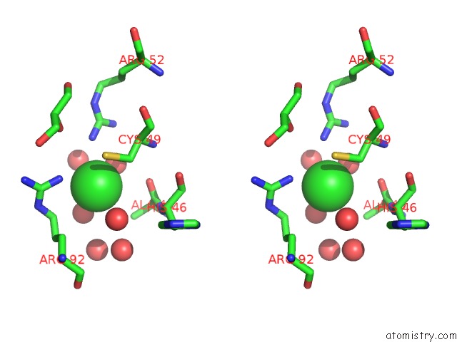
Chlorine binding site 2 out of 5 in 5y5e
Go back to
Chlorine binding site 2 out
of 5 in the Crystal Structure of Phospholipase A2 with Inhibitor
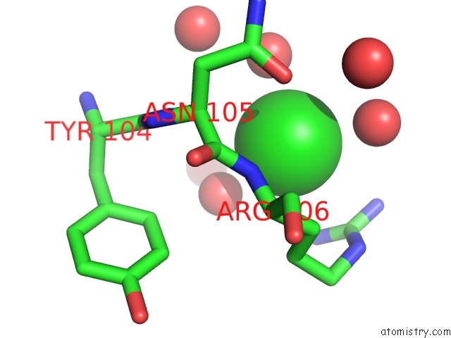
Mono view
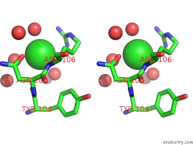
Stereo pair view
Mono view
Stereo pair view
A full contact list of Chlorine with other atoms in the Cl binding
site number 2 of Crystal Structure of Phospholipase A2 with Inhibitor within 5.0Å range:
|
Chlorine binding site 3 out of 5 in 5y5e
Go back to
Chlorine binding site 3 out
of 5 in the Crystal Structure of Phospholipase A2 with Inhibitor
Mono view
Stereo pair view
Mono view
Stereo pair view
A full contact list of Chlorine with other atoms in the Cl binding
site number 3 of Crystal Structure of Phospholipase A2 with Inhibitor within 5.0Å range:
|
Chlorine binding site 4 out of 5 in 5y5e
Go back to
Chlorine binding site 4 out
of 5 in the Crystal Structure of Phospholipase A2 with Inhibitor
Mono view
Stereo pair view
Mono view
Stereo pair view
A full contact list of Chlorine with other atoms in the Cl binding
site number 4 of Crystal Structure of Phospholipase A2 with Inhibitor within 5.0Å range:
|
Chlorine binding site 5 out of 5 in 5y5e
Go back to
Chlorine binding site 5 out
of 5 in the Crystal Structure of Phospholipase A2 with Inhibitor
Mono view
Stereo pair view
Mono view
Stereo pair view
A full contact list of Chlorine with other atoms in the Cl binding
site number 5 of Crystal Structure of Phospholipase A2 with Inhibitor within 5.0Å range:
|
Reference:
S.Hou,
T.Xu,
J.Xu,
L.Qu,
Y.Xu,
L.Chen,
J.Liu.
Structural Basis For Functional Selectivity and Ligand Recognition Revealed By Crystal Structures of Human Secreted Phospholipase A2GROUP Iie Sci Rep V. 7 10815 2017.
ISSN: ESSN 2045-2322
PubMed: 28883454
DOI: 10.1038/S41598-017-11219-8
Page generated: Fri Jul 26 21:17:55 2024
ISSN: ESSN 2045-2322
PubMed: 28883454
DOI: 10.1038/S41598-017-11219-8
Last articles
Zn in 9J0NZn in 9J0O
Zn in 9J0P
Zn in 9FJX
Zn in 9EKB
Zn in 9C0F
Zn in 9CAH
Zn in 9CH0
Zn in 9CH3
Zn in 9CH1