Chlorine »
PDB 5y7x-5yka »
5y9y »
Chlorine in PDB 5y9y: Crystal Structure of the Kdo Hydroxylase Kdoo, A Non-Heme Fe(II) Alphaketoglutarate Dependent Dioxygenase in Complex with Succinate and Co(II)
Protein crystallography data
The structure of Crystal Structure of the Kdo Hydroxylase Kdoo, A Non-Heme Fe(II) Alphaketoglutarate Dependent Dioxygenase in Complex with Succinate and Co(II), PDB code: 5y9y
was solved by
H.S.Chung,
C.W.Pemble,
S.H.Joo,
C.R.Raetz,
with X-Ray Crystallography technique. A brief refinement statistics is given in the table below:
| Resolution Low / High (Å) | 23.57 / 1.60 |
| Space group | P 21 21 21 |
| Cell size a, b, c (Å), α, β, γ (°) | 45.762, 59.316, 116.429, 90.00, 90.00, 90.00 |
| R / Rfree (%) | 15.9 / 19.4 |
Other elements in 5y9y:
The structure of Crystal Structure of the Kdo Hydroxylase Kdoo, A Non-Heme Fe(II) Alphaketoglutarate Dependent Dioxygenase in Complex with Succinate and Co(II) also contains other interesting chemical elements:
| Cobalt | (Co) | 1 atom |
Chlorine Binding Sites:
The binding sites of Chlorine atom in the Crystal Structure of the Kdo Hydroxylase Kdoo, A Non-Heme Fe(II) Alphaketoglutarate Dependent Dioxygenase in Complex with Succinate and Co(II)
(pdb code 5y9y). This binding sites where shown within
5.0 Angstroms radius around Chlorine atom.
In total only one binding site of Chlorine was determined in the Crystal Structure of the Kdo Hydroxylase Kdoo, A Non-Heme Fe(II) Alphaketoglutarate Dependent Dioxygenase in Complex with Succinate and Co(II), PDB code: 5y9y:
In total only one binding site of Chlorine was determined in the Crystal Structure of the Kdo Hydroxylase Kdoo, A Non-Heme Fe(II) Alphaketoglutarate Dependent Dioxygenase in Complex with Succinate and Co(II), PDB code: 5y9y:
Chlorine binding site 1 out of 1 in 5y9y
Go back to
Chlorine binding site 1 out
of 1 in the Crystal Structure of the Kdo Hydroxylase Kdoo, A Non-Heme Fe(II) Alphaketoglutarate Dependent Dioxygenase in Complex with Succinate and Co(II)
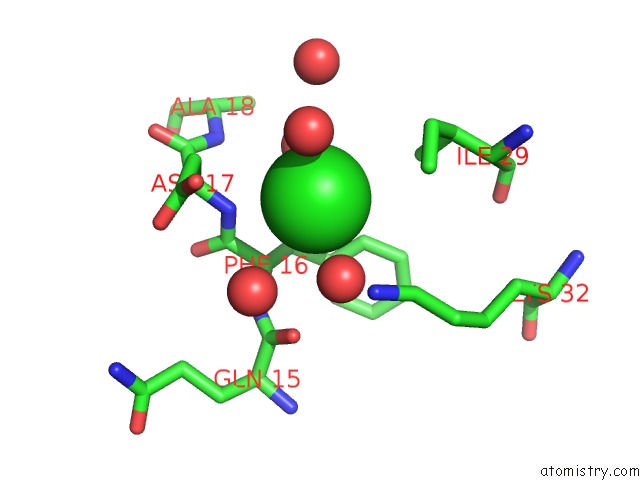
Mono view
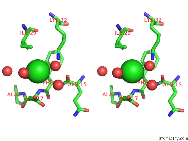
Stereo pair view
Mono view
Stereo pair view
A full contact list of Chlorine with other atoms in the Cl binding
site number 1 of Crystal Structure of the Kdo Hydroxylase Kdoo, A Non-Heme Fe(II) Alphaketoglutarate Dependent Dioxygenase in Complex with Succinate and Co(II) within 5.0Å range:
|
Reference:
H.S.Chung,
C.W.Pemble Iv,
S.H.Joo.
Biochemical and Structural Insights of Fe(II)/Alpha-Ketoglutarate/O2-Dependent Dioxygenase, Kdoo From Methylacidiphilum Infernorum V4 To Be Published.
Page generated: Sat Jul 12 10:54:08 2025
Last articles
Fe in 2YXOFe in 2YRS
Fe in 2YXC
Fe in 2YNM
Fe in 2YVJ
Fe in 2YP1
Fe in 2YU2
Fe in 2YU1
Fe in 2YQB
Fe in 2YOO