Chlorine »
PDB 5y7x-5yka »
5yd2 »
Chlorine in PDB 5yd2: Crystal Structure of Delta 4 Mutant of Ehpsat (Phosphoserine Aminotransferase of Entamoeba Histolytica)
Protein crystallography data
The structure of Crystal Structure of Delta 4 Mutant of Ehpsat (Phosphoserine Aminotransferase of Entamoeba Histolytica), PDB code: 5yd2
was solved by
R.K.Singh,
S.Gourinath,
with X-Ray Crystallography technique. A brief refinement statistics is given in the table below:
| Resolution Low / High (Å) | 50.00 / 2.35 |
| Space group | C 1 2 1 |
| Cell size a, b, c (Å), α, β, γ (°) | 132.238, 67.904, 95.030, 90.00, 110.93, 90.00 |
| R / Rfree (%) | 22.9 / 28.7 |
Chlorine Binding Sites:
The binding sites of Chlorine atom in the Crystal Structure of Delta 4 Mutant of Ehpsat (Phosphoserine Aminotransferase of Entamoeba Histolytica)
(pdb code 5yd2). This binding sites where shown within
5.0 Angstroms radius around Chlorine atom.
In total 2 binding sites of Chlorine where determined in the Crystal Structure of Delta 4 Mutant of Ehpsat (Phosphoserine Aminotransferase of Entamoeba Histolytica), PDB code: 5yd2:
Jump to Chlorine binding site number: 1; 2;
In total 2 binding sites of Chlorine where determined in the Crystal Structure of Delta 4 Mutant of Ehpsat (Phosphoserine Aminotransferase of Entamoeba Histolytica), PDB code: 5yd2:
Jump to Chlorine binding site number: 1; 2;
Chlorine binding site 1 out of 2 in 5yd2
Go back to
Chlorine binding site 1 out
of 2 in the Crystal Structure of Delta 4 Mutant of Ehpsat (Phosphoserine Aminotransferase of Entamoeba Histolytica)
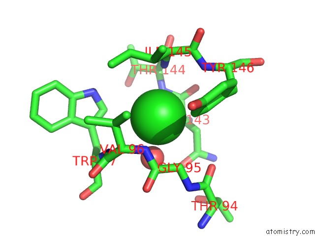
Mono view
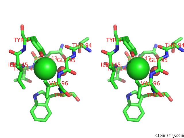
Stereo pair view
Mono view
Stereo pair view
A full contact list of Chlorine with other atoms in the Cl binding
site number 1 of Crystal Structure of Delta 4 Mutant of Ehpsat (Phosphoserine Aminotransferase of Entamoeba Histolytica) within 5.0Å range:
|
Chlorine binding site 2 out of 2 in 5yd2
Go back to
Chlorine binding site 2 out
of 2 in the Crystal Structure of Delta 4 Mutant of Ehpsat (Phosphoserine Aminotransferase of Entamoeba Histolytica)
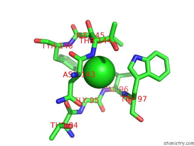
Mono view
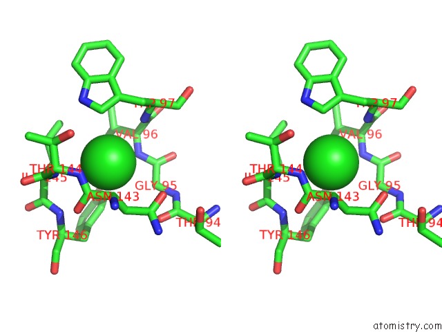
Stereo pair view
Mono view
Stereo pair view
A full contact list of Chlorine with other atoms in the Cl binding
site number 2 of Crystal Structure of Delta 4 Mutant of Ehpsat (Phosphoserine Aminotransferase of Entamoeba Histolytica) within 5.0Å range:
|
Reference:
R.K.Singh,
P.Tomar,
S.Dharavath,
S.Kumar,
S.Gourinath.
N-Terminal Residues Are Crucial For Quaternary Structure and Active Site Conformation For the Phosphoserine Aminotransferase From Enteric Human Parasite E. Histolytica. Int.J.Biol.Macromol. V. 132 1012 2019.
ISSN: ISSN 0141-8130
PubMed: 30959130
DOI: 10.1016/J.IJBIOMAC.2019.04.027
Page generated: Sat Jul 12 10:54:43 2025
ISSN: ISSN 0141-8130
PubMed: 30959130
DOI: 10.1016/J.IJBIOMAC.2019.04.027
Last articles
Fe in 2YXOFe in 2YRS
Fe in 2YXC
Fe in 2YNM
Fe in 2YVJ
Fe in 2YP1
Fe in 2YU2
Fe in 2YU1
Fe in 2YQB
Fe in 2YOO