Chlorine »
PDB 5yke-5z30 »
5yxg »
Chlorine in PDB 5yxg: Crystal Structure of C-Terminal Fragment of Spad From Lactobacillus Rhamnosus Gg Generated By Limited Proteolysis
Protein crystallography data
The structure of Crystal Structure of C-Terminal Fragment of Spad From Lactobacillus Rhamnosus Gg Generated By Limited Proteolysis, PDB code: 5yxg
was solved by
P.Chaurasia,
S.Pratap,
A.Palva,
I.Von Ossowski,
V.Krishnan,
with X-Ray Crystallography technique. A brief refinement statistics is given in the table below:
| Resolution Low / High (Å) | 72.66 / 1.48 |
| Space group | P 21 21 21 |
| Cell size a, b, c (Å), α, β, γ (°) | 50.106, 83.160, 149.403, 90.00, 90.00, 90.00 |
| R / Rfree (%) | 17.7 / 19.5 |
Chlorine Binding Sites:
The binding sites of Chlorine atom in the Crystal Structure of C-Terminal Fragment of Spad From Lactobacillus Rhamnosus Gg Generated By Limited Proteolysis
(pdb code 5yxg). This binding sites where shown within
5.0 Angstroms radius around Chlorine atom.
In total 4 binding sites of Chlorine where determined in the Crystal Structure of C-Terminal Fragment of Spad From Lactobacillus Rhamnosus Gg Generated By Limited Proteolysis, PDB code: 5yxg:
Jump to Chlorine binding site number: 1; 2; 3; 4;
In total 4 binding sites of Chlorine where determined in the Crystal Structure of C-Terminal Fragment of Spad From Lactobacillus Rhamnosus Gg Generated By Limited Proteolysis, PDB code: 5yxg:
Jump to Chlorine binding site number: 1; 2; 3; 4;
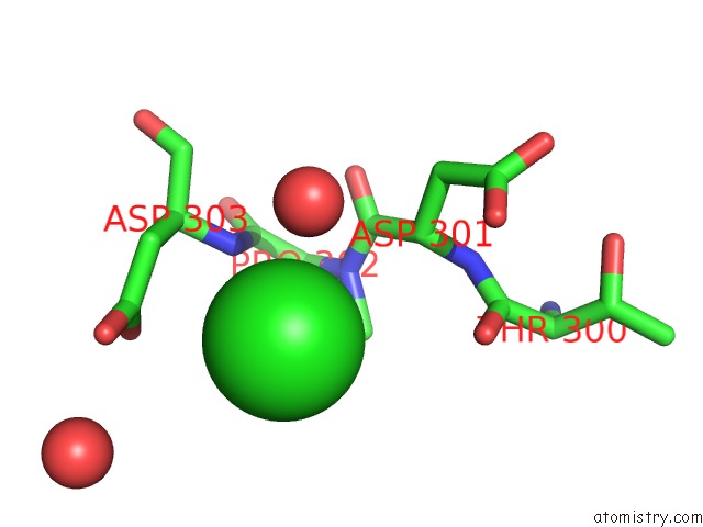
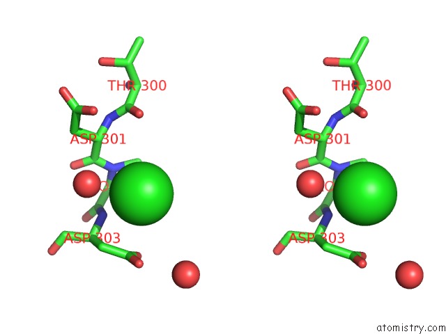
Chlorine binding site 1 out of 4 in 5yxg
Go back to
Chlorine binding site 1 out
of 4 in the Crystal Structure of C-Terminal Fragment of Spad From Lactobacillus Rhamnosus Gg Generated By Limited Proteolysis
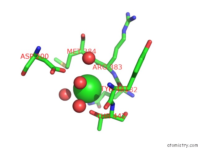
Mono view
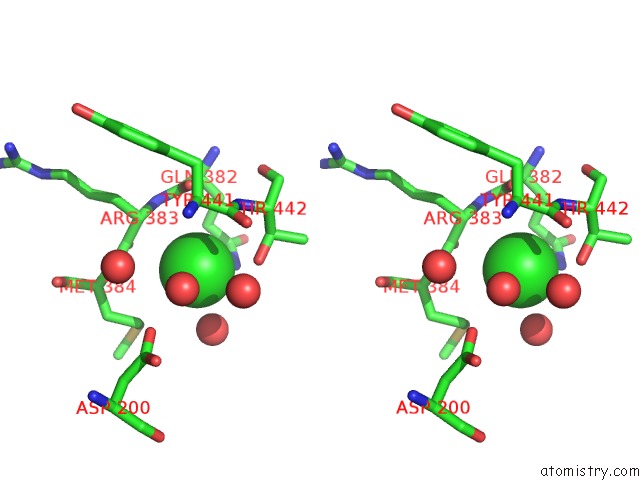
Stereo pair view
Mono view
Stereo pair view
A full contact list of Chlorine with other atoms in the Cl binding
site number 1 of Crystal Structure of C-Terminal Fragment of Spad From Lactobacillus Rhamnosus Gg Generated By Limited Proteolysis within 5.0Å range:
|
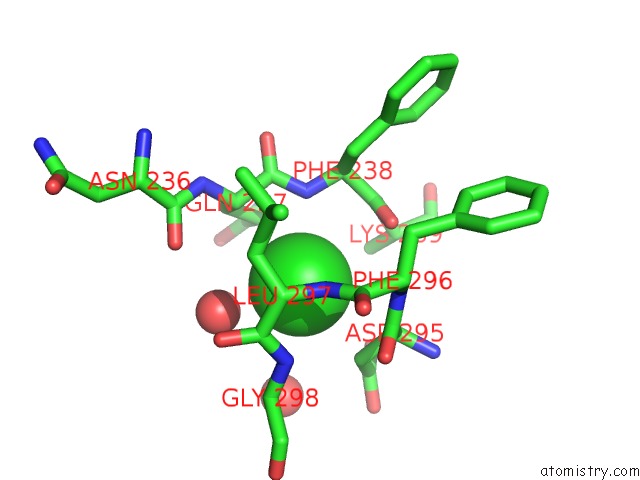
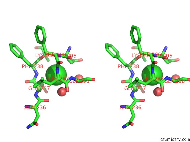
Chlorine binding site 2 out of 4 in 5yxg
Go back to
Chlorine binding site 2 out
of 4 in the Crystal Structure of C-Terminal Fragment of Spad From Lactobacillus Rhamnosus Gg Generated By Limited Proteolysis
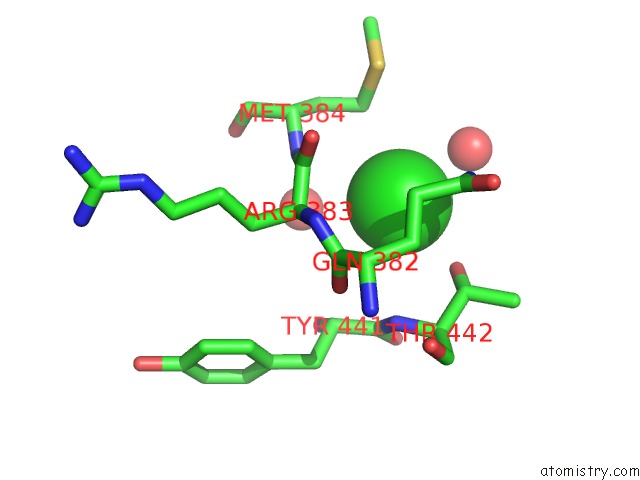
Mono view
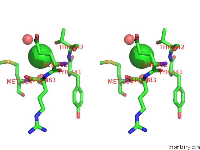
Stereo pair view
Mono view
Stereo pair view
A full contact list of Chlorine with other atoms in the Cl binding
site number 2 of Crystal Structure of C-Terminal Fragment of Spad From Lactobacillus Rhamnosus Gg Generated By Limited Proteolysis within 5.0Å range:
|
Chlorine binding site 3 out of 4 in 5yxg
Go back to
Chlorine binding site 3 out
of 4 in the Crystal Structure of C-Terminal Fragment of Spad From Lactobacillus Rhamnosus Gg Generated By Limited Proteolysis
Mono view
Stereo pair view
Mono view
Stereo pair view
A full contact list of Chlorine with other atoms in the Cl binding
site number 3 of Crystal Structure of C-Terminal Fragment of Spad From Lactobacillus Rhamnosus Gg Generated By Limited Proteolysis within 5.0Å range:
|
Chlorine binding site 4 out of 4 in 5yxg
Go back to
Chlorine binding site 4 out
of 4 in the Crystal Structure of C-Terminal Fragment of Spad From Lactobacillus Rhamnosus Gg Generated By Limited Proteolysis
Mono view
Stereo pair view
Mono view
Stereo pair view
A full contact list of Chlorine with other atoms in the Cl binding
site number 4 of Crystal Structure of C-Terminal Fragment of Spad From Lactobacillus Rhamnosus Gg Generated By Limited Proteolysis within 5.0Å range:
|
Reference:
P.Chaurasia,
S.Pratap,
A.Palva,
I.Von Ossowski,
V.Krishnan.
Bent Conformation of A Backbone Pilin N-Terminal Domain Supports A Three-Stage Pilus Assembly Mechanism. Commun Biol V. 1 94 2018.
ISSN: ESSN 2399-3642
PubMed: 30271975
DOI: 10.1038/S42003-018-0100-0
Page generated: Sat Jul 12 11:04:53 2025
ISSN: ESSN 2399-3642
PubMed: 30271975
DOI: 10.1038/S42003-018-0100-0
Last articles
Fe in 2YXOFe in 2YRS
Fe in 2YXC
Fe in 2YNM
Fe in 2YVJ
Fe in 2YP1
Fe in 2YU2
Fe in 2YU1
Fe in 2YQB
Fe in 2YOO