Chlorine »
PDB 6a18-6aek »
6a6i »
Chlorine in PDB 6a6i: Crystal Structure of the Winged-Helix Domain of Cockayne Syndrome Group B Protein in Complex with Ubiquitin
Protein crystallography data
The structure of Crystal Structure of the Winged-Helix Domain of Cockayne Syndrome Group B Protein in Complex with Ubiquitin, PDB code: 6a6i
was solved by
T.S.Takahashi,
Y.Sato,
S.Fukai,
with X-Ray Crystallography technique. A brief refinement statistics is given in the table below:
| Resolution Low / High (Å) | 39.96 / 2.60 |
| Space group | P 1 21 1 |
| Cell size a, b, c (Å), α, β, γ (°) | 54.161, 101.705, 66.008, 90.00, 101.81, 90.00 |
| R / Rfree (%) | 17.9 / 23.6 |
Chlorine Binding Sites:
The binding sites of Chlorine atom in the Crystal Structure of the Winged-Helix Domain of Cockayne Syndrome Group B Protein in Complex with Ubiquitin
(pdb code 6a6i). This binding sites where shown within
5.0 Angstroms radius around Chlorine atom.
In total only one binding site of Chlorine was determined in the Crystal Structure of the Winged-Helix Domain of Cockayne Syndrome Group B Protein in Complex with Ubiquitin, PDB code: 6a6i:
In total only one binding site of Chlorine was determined in the Crystal Structure of the Winged-Helix Domain of Cockayne Syndrome Group B Protein in Complex with Ubiquitin, PDB code: 6a6i:
Chlorine binding site 1 out of 1 in 6a6i
Go back to
Chlorine binding site 1 out
of 1 in the Crystal Structure of the Winged-Helix Domain of Cockayne Syndrome Group B Protein in Complex with Ubiquitin
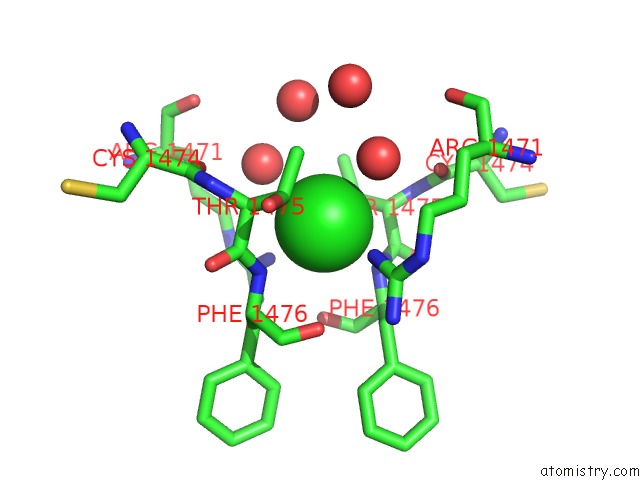
Mono view
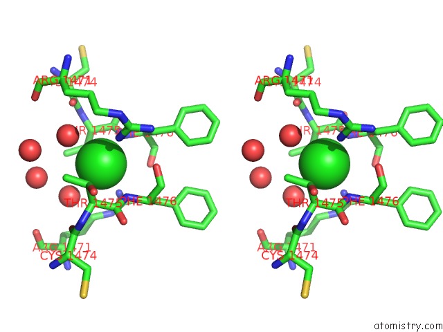
Stereo pair view
Mono view
Stereo pair view
A full contact list of Chlorine with other atoms in the Cl binding
site number 1 of Crystal Structure of the Winged-Helix Domain of Cockayne Syndrome Group B Protein in Complex with Ubiquitin within 5.0Å range:
|
Reference:
T.S.Takahashi,
Y.Sato,
A.Yamagata,
S.Goto-Ito,
M.Saijo,
S.Fukai.
Structural Basis of Ubiquitin Recognition By the Winged-Helix Domain of Cockayne Syndrome Group B Protein. Nucleic Acids Res. V. 47 3784 2019.
ISSN: ESSN 1362-4962
PubMed: 30753618
DOI: 10.1093/NAR/GKZ081
Page generated: Fri Jul 26 21:55:14 2024
ISSN: ESSN 1362-4962
PubMed: 30753618
DOI: 10.1093/NAR/GKZ081
Last articles
Zn in 9JYWZn in 9IR4
Zn in 9IR3
Zn in 9GMX
Zn in 9GMW
Zn in 9JEJ
Zn in 9ERF
Zn in 9ERE
Zn in 9EGV
Zn in 9EGW