Chlorine »
PDB 6aqe-6axv »
6aw1 »
Chlorine in PDB 6aw1: Crystal Structure of CEACAM3
Protein crystallography data
The structure of Crystal Structure of CEACAM3, PDB code: 6aw1
was solved by
D.A.Bonsor,
E.J.Sundberg,
with X-Ray Crystallography technique. A brief refinement statistics is given in the table below:
| Resolution Low / High (Å) | 80.46 / 2.10 |
| Space group | I 41 2 2 |
| Cell size a, b, c (Å), α, β, γ (°) | 94.433, 94.433, 153.707, 90.00, 90.00, 90.00 |
| R / Rfree (%) | 20.5 / 22.3 |
Chlorine Binding Sites:
The binding sites of Chlorine atom in the Crystal Structure of CEACAM3
(pdb code 6aw1). This binding sites where shown within
5.0 Angstroms radius around Chlorine atom.
In total only one binding site of Chlorine was determined in the Crystal Structure of CEACAM3, PDB code: 6aw1:
In total only one binding site of Chlorine was determined in the Crystal Structure of CEACAM3, PDB code: 6aw1:
Chlorine binding site 1 out of 1 in 6aw1
Go back to
Chlorine binding site 1 out
of 1 in the Crystal Structure of CEACAM3
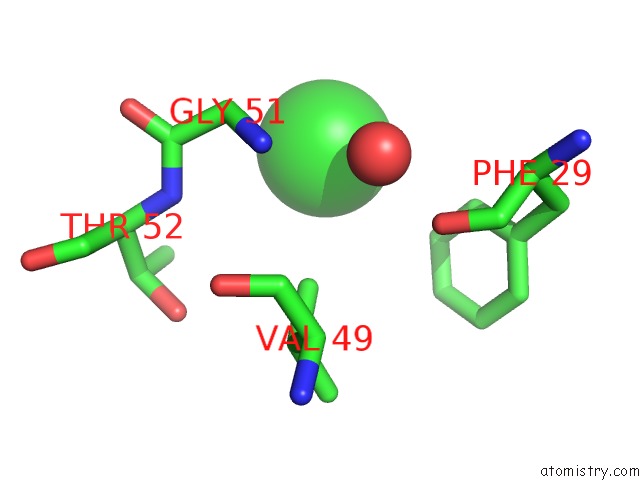
Mono view
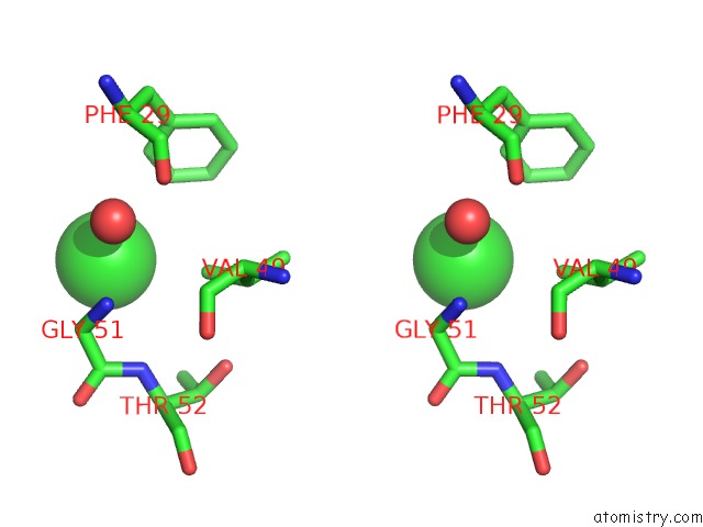
Stereo pair view
Mono view
Stereo pair view
A full contact list of Chlorine with other atoms in the Cl binding
site number 1 of Crystal Structure of CEACAM3 within 5.0Å range:
|
Reference:
D.A.Bonsor,
Q.Zhao,
B.Schmidinger,
E.Weiss,
J.Wang,
D.Deredge,
R.Beadenkopf,
B.Dow,
W.Fischer,
D.Beckett,
P.L.Wintrode,
R.Haas,
E.J.Sundberg.
Thehelicobacter Pyloriadhesin Protein Hopq Exploits the Dimer Interface of Human Ceacams to Facilitate Translocation of the Oncoprotein Caga. Embo J. V. 37 2018.
ISSN: ESSN 1460-2075
PubMed: 29724755
DOI: 10.15252/EMBJ.201798664
Page generated: Fri Jul 26 22:19:08 2024
ISSN: ESSN 1460-2075
PubMed: 29724755
DOI: 10.15252/EMBJ.201798664
Last articles
Zn in 9J0NZn in 9J0O
Zn in 9J0P
Zn in 9FJX
Zn in 9EKB
Zn in 9C0F
Zn in 9CAH
Zn in 9CH0
Zn in 9CH3
Zn in 9CH1