Chlorine »
PDB 6aqe-6axv »
6aw5 »
Chlorine in PDB 6aw5: 1.90A Resolution Structure of Catechol O-Methyltransferase (Comt) L136M (Hexagonal Form) From Nannospalax Galili
Protein crystallography data
The structure of 1.90A Resolution Structure of Catechol O-Methyltransferase (Comt) L136M (Hexagonal Form) From Nannospalax Galili, PDB code: 6aw5
was solved by
S.Lovell,
N.Mehzabeen,
K.P.Battaile,
Y.Deng,
R.P.Hanzlik,
I.Shams,
J.Moskovitz,
with X-Ray Crystallography technique. A brief refinement statistics is given in the table below:
| Resolution Low / High (Å) | 36.14 / 1.90 |
| Space group | P 62 |
| Cell size a, b, c (Å), α, β, γ (°) | 95.186, 95.186, 75.172, 90.00, 90.00, 120.00 |
| R / Rfree (%) | 16.3 / 19.2 |
Other elements in 6aw5:
The structure of 1.90A Resolution Structure of Catechol O-Methyltransferase (Comt) L136M (Hexagonal Form) From Nannospalax Galili also contains other interesting chemical elements:
| Sodium | (Na) | 1 atom |
Chlorine Binding Sites:
The binding sites of Chlorine atom in the 1.90A Resolution Structure of Catechol O-Methyltransferase (Comt) L136M (Hexagonal Form) From Nannospalax Galili
(pdb code 6aw5). This binding sites where shown within
5.0 Angstroms radius around Chlorine atom.
In total 2 binding sites of Chlorine where determined in the 1.90A Resolution Structure of Catechol O-Methyltransferase (Comt) L136M (Hexagonal Form) From Nannospalax Galili, PDB code: 6aw5:
Jump to Chlorine binding site number: 1; 2;
In total 2 binding sites of Chlorine where determined in the 1.90A Resolution Structure of Catechol O-Methyltransferase (Comt) L136M (Hexagonal Form) From Nannospalax Galili, PDB code: 6aw5:
Jump to Chlorine binding site number: 1; 2;
Chlorine binding site 1 out of 2 in 6aw5
Go back to
Chlorine binding site 1 out
of 2 in the 1.90A Resolution Structure of Catechol O-Methyltransferase (Comt) L136M (Hexagonal Form) From Nannospalax Galili
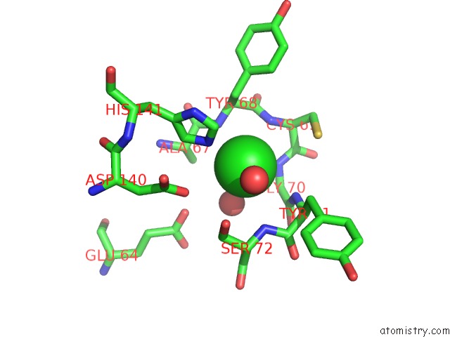
Mono view
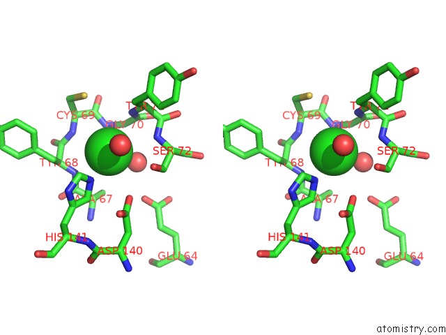
Stereo pair view
Mono view
Stereo pair view
A full contact list of Chlorine with other atoms in the Cl binding
site number 1 of 1.90A Resolution Structure of Catechol O-Methyltransferase (Comt) L136M (Hexagonal Form) From Nannospalax Galili within 5.0Å range:
|
Chlorine binding site 2 out of 2 in 6aw5
Go back to
Chlorine binding site 2 out
of 2 in the 1.90A Resolution Structure of Catechol O-Methyltransferase (Comt) L136M (Hexagonal Form) From Nannospalax Galili
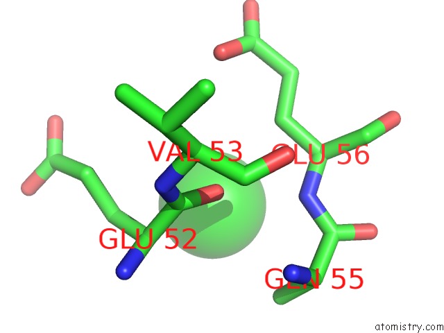
Mono view
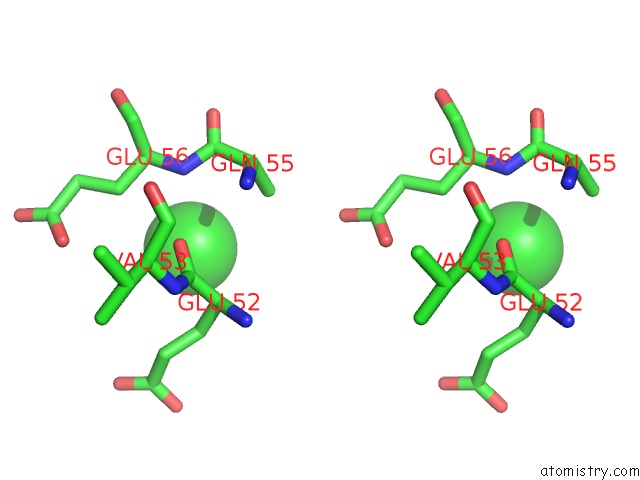
Stereo pair view
Mono view
Stereo pair view
A full contact list of Chlorine with other atoms in the Cl binding
site number 2 of 1.90A Resolution Structure of Catechol O-Methyltransferase (Comt) L136M (Hexagonal Form) From Nannospalax Galili within 5.0Å range:
|
Reference:
Y.Deng,
S.Lovell,
N.Mehzabeen,
K.P.Battaile,
R.P.Hanzlik,
I.Shams,
J.Moskovitz.
Crystal Structure of the Catechol-O-Methyl Transferase (Comt) Enzyme of the Subterranean Mole Rat (Spalax) and the Effect of L136M Substitution To Be Published.
Page generated: Fri Jul 26 22:19:18 2024
Last articles
Zn in 9J0NZn in 9J0O
Zn in 9J0P
Zn in 9FJX
Zn in 9EKB
Zn in 9C0F
Zn in 9CAH
Zn in 9CH0
Zn in 9CH3
Zn in 9CH1