Chlorine »
PDB 6b4n-6bb2 »
6b5e »
Chlorine in PDB 6b5e: Mycobacterium Tuberculosis Rmla in Complex with Dtdp-Glucose
Enzymatic activity of Mycobacterium Tuberculosis Rmla in Complex with Dtdp-Glucose
All present enzymatic activity of Mycobacterium Tuberculosis Rmla in Complex with Dtdp-Glucose:
2.7.7.24;
2.7.7.24;
Protein crystallography data
The structure of Mycobacterium Tuberculosis Rmla in Complex with Dtdp-Glucose, PDB code: 6b5e
was solved by
H.A.Brown,
H.M.Holden,
with X-Ray Crystallography technique. A brief refinement statistics is given in the table below:
| Resolution Low / High (Å) | 50.00 / 1.85 |
| Space group | P 21 21 21 |
| Cell size a, b, c (Å), α, β, γ (°) | 72.323, 111.264, 290.363, 90.00, 90.00, 90.00 |
| R / Rfree (%) | 17.1 / 21.8 |
Other elements in 6b5e:
The structure of Mycobacterium Tuberculosis Rmla in Complex with Dtdp-Glucose also contains other interesting chemical elements:
| Magnesium | (Mg) | 9 atoms |
| Sodium | (Na) | 2 atoms |
Chlorine Binding Sites:
The binding sites of Chlorine atom in the Mycobacterium Tuberculosis Rmla in Complex with Dtdp-Glucose
(pdb code 6b5e). This binding sites where shown within
5.0 Angstroms radius around Chlorine atom.
In total only one binding site of Chlorine was determined in the Mycobacterium Tuberculosis Rmla in Complex with Dtdp-Glucose, PDB code: 6b5e:
In total only one binding site of Chlorine was determined in the Mycobacterium Tuberculosis Rmla in Complex with Dtdp-Glucose, PDB code: 6b5e:
Chlorine binding site 1 out of 1 in 6b5e
Go back to
Chlorine binding site 1 out
of 1 in the Mycobacterium Tuberculosis Rmla in Complex with Dtdp-Glucose
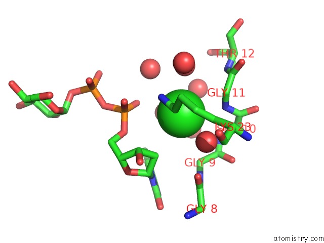
Mono view
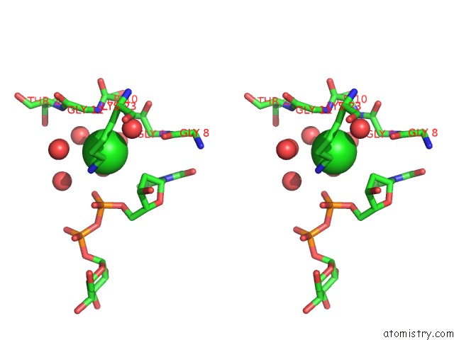
Stereo pair view
Mono view
Stereo pair view
A full contact list of Chlorine with other atoms in the Cl binding
site number 1 of Mycobacterium Tuberculosis Rmla in Complex with Dtdp-Glucose within 5.0Å range:
|
Reference:
H.A.Brown,
J.B.Thoden,
P.A.Tipton,
H.M.Holden.
The Structure of Glucose-1-Phosphate Thymidylyltransferase From Mycobacterium Tuberculosis Reveals the Location of An Essential Magnesium Ion in the Rmla-Type Enzymes. Protein Sci. V. 27 441 2018.
ISSN: ESSN 1469-896X
PubMed: 29076563
DOI: 10.1002/PRO.3333
Page generated: Fri Jul 26 22:33:32 2024
ISSN: ESSN 1469-896X
PubMed: 29076563
DOI: 10.1002/PRO.3333
Last articles
Zn in 9J0NZn in 9J0O
Zn in 9J0P
Zn in 9FJX
Zn in 9EKB
Zn in 9C0F
Zn in 9CAH
Zn in 9CH0
Zn in 9CH3
Zn in 9CH1