Chlorine »
PDB 6bb3-6bmc »
6bel »
Chlorine in PDB 6bel: Ternary Complex Crystal Structure of Dna Polymerase Beta with R-Isomer of Beta-Gamma-Chf-Dctp
Enzymatic activity of Ternary Complex Crystal Structure of Dna Polymerase Beta with R-Isomer of Beta-Gamma-Chf-Dctp
All present enzymatic activity of Ternary Complex Crystal Structure of Dna Polymerase Beta with R-Isomer of Beta-Gamma-Chf-Dctp:
2.7.7.7;
2.7.7.7;
Protein crystallography data
The structure of Ternary Complex Crystal Structure of Dna Polymerase Beta with R-Isomer of Beta-Gamma-Chf-Dctp, PDB code: 6bel
was solved by
V.K.Batra,
S.H.Wilson,
with X-Ray Crystallography technique. A brief refinement statistics is given in the table below:
| Resolution Low / High (Å) | 23.70 / 1.90 |
| Space group | P 1 21 1 |
| Cell size a, b, c (Å), α, β, γ (°) | 50.709, 79.533, 55.458, 90.00, 107.66, 90.00 |
| R / Rfree (%) | 16.8 / 20.2 |
Other elements in 6bel:
The structure of Ternary Complex Crystal Structure of Dna Polymerase Beta with R-Isomer of Beta-Gamma-Chf-Dctp also contains other interesting chemical elements:
| Fluorine | (F) | 1 atom |
| Magnesium | (Mg) | 2 atoms |
| Sodium | (Na) | 3 atoms |
Chlorine Binding Sites:
The binding sites of Chlorine atom in the Ternary Complex Crystal Structure of Dna Polymerase Beta with R-Isomer of Beta-Gamma-Chf-Dctp
(pdb code 6bel). This binding sites where shown within
5.0 Angstroms radius around Chlorine atom.
In total only one binding site of Chlorine was determined in the Ternary Complex Crystal Structure of Dna Polymerase Beta with R-Isomer of Beta-Gamma-Chf-Dctp, PDB code: 6bel:
In total only one binding site of Chlorine was determined in the Ternary Complex Crystal Structure of Dna Polymerase Beta with R-Isomer of Beta-Gamma-Chf-Dctp, PDB code: 6bel:
Chlorine binding site 1 out of 1 in 6bel
Go back to
Chlorine binding site 1 out
of 1 in the Ternary Complex Crystal Structure of Dna Polymerase Beta with R-Isomer of Beta-Gamma-Chf-Dctp
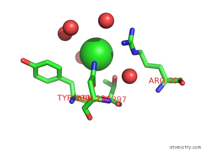
Mono view
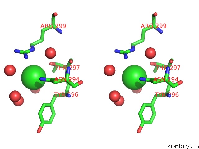
Stereo pair view
Mono view
Stereo pair view
A full contact list of Chlorine with other atoms in the Cl binding
site number 1 of Ternary Complex Crystal Structure of Dna Polymerase Beta with R-Isomer of Beta-Gamma-Chf-Dctp within 5.0Å range:
|
Reference:
V.K.Batra,
K.Oertell,
W.A.Beard,
B.A.Kashemirov,
C.E.Mckenna,
M.F.Goodman,
S.H.Wilson.
Mapping Functional Substrate-Enzyme Interactions in the Pol Beta Active Site Through Chemical Biology: Structural Responses to Acidity Modification of Incoming Dntps. Biochemistry V. 57 3934 2018.
ISSN: ISSN 1520-4995
PubMed: 29874056
DOI: 10.1021/ACS.BIOCHEM.8B00418
Page generated: Fri Jul 26 22:46:07 2024
ISSN: ISSN 1520-4995
PubMed: 29874056
DOI: 10.1021/ACS.BIOCHEM.8B00418
Last articles
Zn in 9J0NZn in 9J0O
Zn in 9J0P
Zn in 9FJX
Zn in 9EKB
Zn in 9C0F
Zn in 9CAH
Zn in 9CH0
Zn in 9CH3
Zn in 9CH1