Chlorine »
PDB 6bmr-6bry »
6bq8 »
Chlorine in PDB 6bq8: Joint X-Ray/Neutron Structure of Pkg II Cnb-B Domain in Complex with 8-Pcpt-Cgmp
Enzymatic activity of Joint X-Ray/Neutron Structure of Pkg II Cnb-B Domain in Complex with 8-Pcpt-Cgmp
All present enzymatic activity of Joint X-Ray/Neutron Structure of Pkg II Cnb-B Domain in Complex with 8-Pcpt-Cgmp:
2.7.11.12;
2.7.11.12;
Protein crystallography data
The structure of Joint X-Ray/Neutron Structure of Pkg II Cnb-B Domain in Complex with 8-Pcpt-Cgmp, PDB code: 6bq8
was solved by
C.Kim,
A.Kovalevsky,
O.Gerlits,
with X-Ray Crystallography technique. A brief refinement statistics is given in the table below:
| Resolution Low / High (Å) | N/A / 2.00 |
| Space group | P 21 21 21 |
| Cell size a, b, c (Å), α, β, γ (°) | 43.650, 51.300, 68.200, 90.00, 90.00, 90.00 |
| R / Rfree (%) | 23.2 / 28.8 |
Other elements in 6bq8:
The structure of Joint X-Ray/Neutron Structure of Pkg II Cnb-B Domain in Complex with 8-Pcpt-Cgmp also contains other interesting chemical elements:
| Strontium | (Sr) | 1 atom |
Chlorine Binding Sites:
The binding sites of Chlorine atom in the Joint X-Ray/Neutron Structure of Pkg II Cnb-B Domain in Complex with 8-Pcpt-Cgmp
(pdb code 6bq8). This binding sites where shown within
5.0 Angstroms radius around Chlorine atom.
In total only one binding site of Chlorine was determined in the Joint X-Ray/Neutron Structure of Pkg II Cnb-B Domain in Complex with 8-Pcpt-Cgmp, PDB code: 6bq8:
In total only one binding site of Chlorine was determined in the Joint X-Ray/Neutron Structure of Pkg II Cnb-B Domain in Complex with 8-Pcpt-Cgmp, PDB code: 6bq8:
Chlorine binding site 1 out of 1 in 6bq8
Go back to
Chlorine binding site 1 out
of 1 in the Joint X-Ray/Neutron Structure of Pkg II Cnb-B Domain in Complex with 8-Pcpt-Cgmp
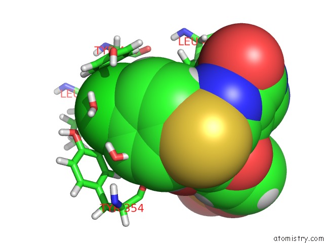
Mono view
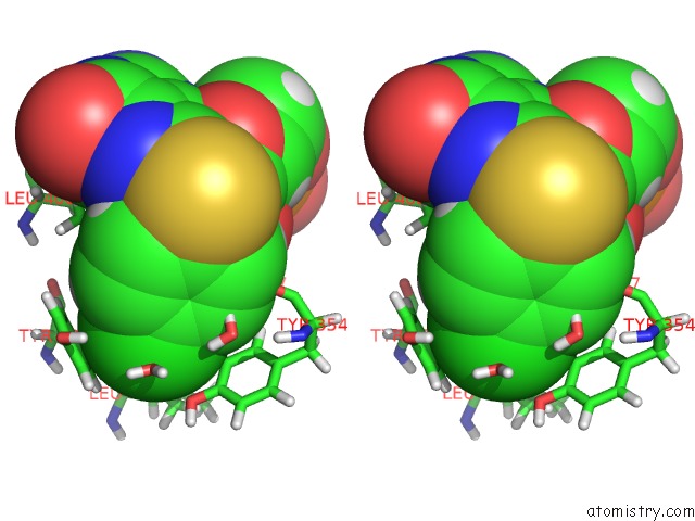
Stereo pair view
Mono view
Stereo pair view
A full contact list of Chlorine with other atoms in the Cl binding
site number 1 of Joint X-Ray/Neutron Structure of Pkg II Cnb-B Domain in Complex with 8-Pcpt-Cgmp within 5.0Å range:
|
Reference:
O.Gerlits,
J.C.Campbell,
M.P.Blakeley,
C.Kim,
A.Kovalevsky.
Neutron Crystallography Detects Differences in Protein Dynamics: Structure of the Pkg II Cyclic Nucleotide Binding Domain in Complex with An Activator. Biochemistry V. 57 1833 2018.
ISSN: ISSN 1520-4995
PubMed: 29517905
DOI: 10.1021/ACS.BIOCHEM.8B00010
Page generated: Fri Jul 26 23:00:34 2024
ISSN: ISSN 1520-4995
PubMed: 29517905
DOI: 10.1021/ACS.BIOCHEM.8B00010
Last articles
Zn in 9J0NZn in 9J0O
Zn in 9J0P
Zn in 9FJX
Zn in 9EKB
Zn in 9C0F
Zn in 9CAH
Zn in 9CH0
Zn in 9CH3
Zn in 9CH1