Chlorine »
PDB 6bsd-6bza »
6bvq »
Chlorine in PDB 6bvq: Crystal Structure of 3-Hydroxyanthranilate-3,4-Dioxygenase N27A From Cupriavidus Metallidurans in Complex with 4-Cl-3-Haa
Enzymatic activity of Crystal Structure of 3-Hydroxyanthranilate-3,4-Dioxygenase N27A From Cupriavidus Metallidurans in Complex with 4-Cl-3-Haa
All present enzymatic activity of Crystal Structure of 3-Hydroxyanthranilate-3,4-Dioxygenase N27A From Cupriavidus Metallidurans in Complex with 4-Cl-3-Haa:
1.13.11.6;
1.13.11.6;
Protein crystallography data
The structure of Crystal Structure of 3-Hydroxyanthranilate-3,4-Dioxygenase N27A From Cupriavidus Metallidurans in Complex with 4-Cl-3-Haa, PDB code: 6bvq
was solved by
Y.Yang,
F.Liu,
A.Liu,
with X-Ray Crystallography technique. A brief refinement statistics is given in the table below:
| Resolution Low / High (Å) | 31.12 / 2.08 |
| Space group | P 65 2 2 |
| Cell size a, b, c (Å), α, β, γ (°) | 58.608, 58.608, 236.316, 90.00, 90.00, 120.00 |
| R / Rfree (%) | 22.8 / 26.7 |
Other elements in 6bvq:
The structure of Crystal Structure of 3-Hydroxyanthranilate-3,4-Dioxygenase N27A From Cupriavidus Metallidurans in Complex with 4-Cl-3-Haa also contains other interesting chemical elements:
| Iron | (Fe) | 2 atoms |
Chlorine Binding Sites:
The binding sites of Chlorine atom in the Crystal Structure of 3-Hydroxyanthranilate-3,4-Dioxygenase N27A From Cupriavidus Metallidurans in Complex with 4-Cl-3-Haa
(pdb code 6bvq). This binding sites where shown within
5.0 Angstroms radius around Chlorine atom.
In total only one binding site of Chlorine was determined in the Crystal Structure of 3-Hydroxyanthranilate-3,4-Dioxygenase N27A From Cupriavidus Metallidurans in Complex with 4-Cl-3-Haa, PDB code: 6bvq:
In total only one binding site of Chlorine was determined in the Crystal Structure of 3-Hydroxyanthranilate-3,4-Dioxygenase N27A From Cupriavidus Metallidurans in Complex with 4-Cl-3-Haa, PDB code: 6bvq:
Chlorine binding site 1 out of 1 in 6bvq
Go back to
Chlorine binding site 1 out
of 1 in the Crystal Structure of 3-Hydroxyanthranilate-3,4-Dioxygenase N27A From Cupriavidus Metallidurans in Complex with 4-Cl-3-Haa
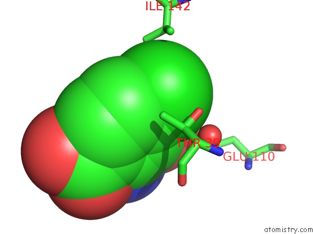
Mono view
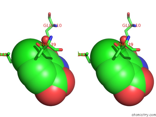
Stereo pair view
Mono view
Stereo pair view
A full contact list of Chlorine with other atoms in the Cl binding
site number 1 of Crystal Structure of 3-Hydroxyanthranilate-3,4-Dioxygenase N27A From Cupriavidus Metallidurans in Complex with 4-Cl-3-Haa within 5.0Å range:
|
Reference:
Y.Yang,
F.Liu,
A.Liu.
Adapting to Oxygen: 3-Hydroxyanthrinilate 3,4-Dioxygenase Employs Loop Dynamics to Accommodate Two Substrates with Disparate Polarities. J. Biol. Chem. V. 293 10415 2018.
ISSN: ESSN 1083-351X
PubMed: 29784877
DOI: 10.1074/JBC.RA118.002698
Page generated: Fri Jul 26 23:08:55 2024
ISSN: ESSN 1083-351X
PubMed: 29784877
DOI: 10.1074/JBC.RA118.002698
Last articles
Zn in 9MJ5Zn in 9HNW
Zn in 9G0L
Zn in 9FNE
Zn in 9DZN
Zn in 9E0I
Zn in 9D32
Zn in 9DAK
Zn in 8ZXC
Zn in 8ZUF