Chlorine »
PDB 6c7i-6cdq »
6cbl »
Chlorine in PDB 6cbl: X-Ray Structure of Neob From Streptomyces Fradiae in Complex with Neamine As An External Aldimine
Enzymatic activity of X-Ray Structure of Neob From Streptomyces Fradiae in Complex with Neamine As An External Aldimine
All present enzymatic activity of X-Ray Structure of Neob From Streptomyces Fradiae in Complex with Neamine As An External Aldimine:
2.6.1.93; 2.6.1.95;
2.6.1.93; 2.6.1.95;
Protein crystallography data
The structure of X-Ray Structure of Neob From Streptomyces Fradiae in Complex with Neamine As An External Aldimine, PDB code: 6cbl
was solved by
J.B.Thoden,
G.T.Dow,
H.M.Holden,
with X-Ray Crystallography technique. A brief refinement statistics is given in the table below:
| Resolution Low / High (Å) | 30.00 / 1.60 |
| Space group | P 1 21 1 |
| Cell size a, b, c (Å), α, β, γ (°) | 70.363, 107.363, 217.528, 90.00, 98.24, 90.00 |
| R / Rfree (%) | 19.6 / 24.3 |
Chlorine Binding Sites:
The binding sites of Chlorine atom in the X-Ray Structure of Neob From Streptomyces Fradiae in Complex with Neamine As An External Aldimine
(pdb code 6cbl). This binding sites where shown within
5.0 Angstroms radius around Chlorine atom.
In total only one binding site of Chlorine was determined in the X-Ray Structure of Neob From Streptomyces Fradiae in Complex with Neamine As An External Aldimine, PDB code: 6cbl:
In total only one binding site of Chlorine was determined in the X-Ray Structure of Neob From Streptomyces Fradiae in Complex with Neamine As An External Aldimine, PDB code: 6cbl:
Chlorine binding site 1 out of 1 in 6cbl
Go back to
Chlorine binding site 1 out
of 1 in the X-Ray Structure of Neob From Streptomyces Fradiae in Complex with Neamine As An External Aldimine
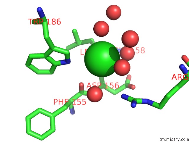
Mono view
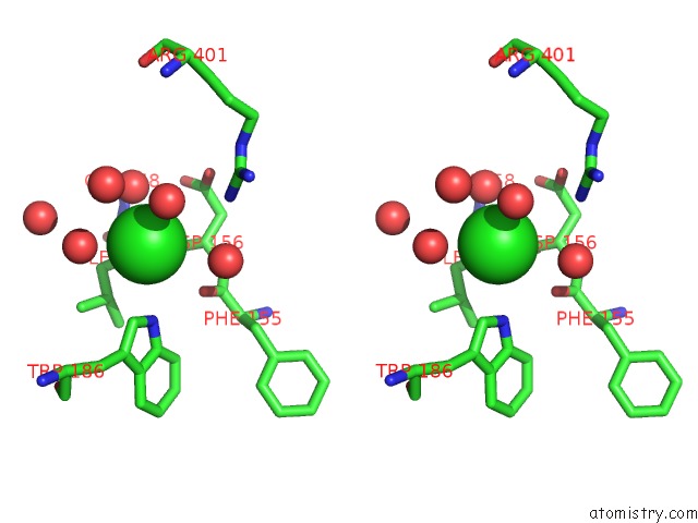
Stereo pair view
Mono view
Stereo pair view
A full contact list of Chlorine with other atoms in the Cl binding
site number 1 of X-Ray Structure of Neob From Streptomyces Fradiae in Complex with Neamine As An External Aldimine within 5.0Å range:
|
Reference:
G.T.Dow,
J.B.Thoden,
H.M.Holden.
The Three-Dimensional Structure of Neob: An Aminotransferase Involved in the Biosynthesis of Neomycin. Protein Sci. V. 27 945 2018.
ISSN: ESSN 1469-896X
PubMed: 29516565
DOI: 10.1002/PRO.3400
Page generated: Fri Jul 26 23:24:51 2024
ISSN: ESSN 1469-896X
PubMed: 29516565
DOI: 10.1002/PRO.3400
Last articles
Zn in 9J0NZn in 9J0O
Zn in 9J0P
Zn in 9FJX
Zn in 9EKB
Zn in 9C0F
Zn in 9CAH
Zn in 9CH0
Zn in 9CH3
Zn in 9CH1