Chlorine »
PDB 6cdr-6cod »
6cn8 »
Chlorine in PDB 6cn8: High-Resolution Structure of CLPC1-Ntd Binding to Rufomycin-I
Protein crystallography data
The structure of High-Resolution Structure of CLPC1-Ntd Binding to Rufomycin-I, PDB code: 6cn8
was solved by
C.Abad-Zapatero,
N.W.Wolf,
with X-Ray Crystallography technique. A brief refinement statistics is given in the table below:
| Resolution Low / High (Å) | 19.40 / 1.40 |
| Space group | P 41 3 2 |
| Cell size a, b, c (Å), α, β, γ (°) | 109.758, 109.758, 109.758, 90.00, 90.00, 90.00 |
| R / Rfree (%) | 15.9 / 18.5 |
Other elements in 6cn8:
The structure of High-Resolution Structure of CLPC1-Ntd Binding to Rufomycin-I also contains other interesting chemical elements:
| Sodium | (Na) | 4 atoms |
Chlorine Binding Sites:
The binding sites of Chlorine atom in the High-Resolution Structure of CLPC1-Ntd Binding to Rufomycin-I
(pdb code 6cn8). This binding sites where shown within
5.0 Angstroms radius around Chlorine atom.
In total 4 binding sites of Chlorine where determined in the High-Resolution Structure of CLPC1-Ntd Binding to Rufomycin-I, PDB code: 6cn8:
Jump to Chlorine binding site number: 1; 2; 3; 4;
In total 4 binding sites of Chlorine where determined in the High-Resolution Structure of CLPC1-Ntd Binding to Rufomycin-I, PDB code: 6cn8:
Jump to Chlorine binding site number: 1; 2; 3; 4;
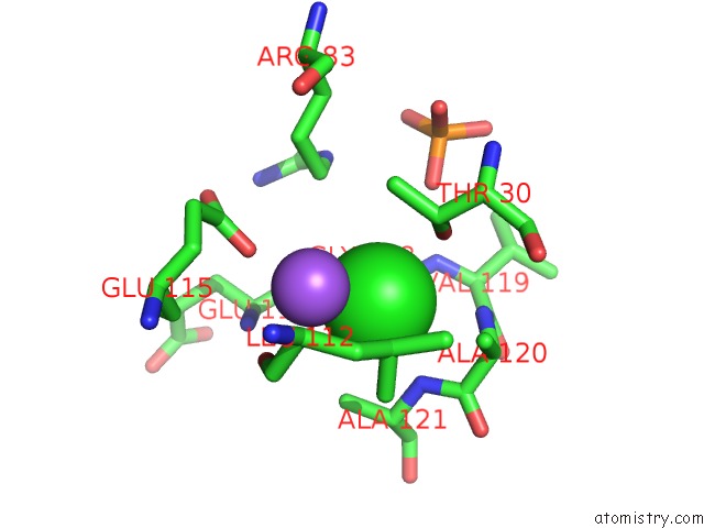
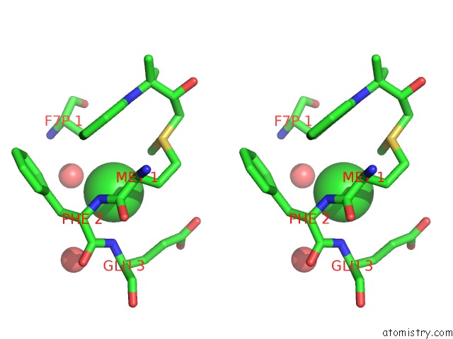
Chlorine binding site 1 out of 4 in 6cn8
Go back to
Chlorine binding site 1 out
of 4 in the High-Resolution Structure of CLPC1-Ntd Binding to Rufomycin-I
Mono view

Stereo pair view
Mono view
Stereo pair view
A full contact list of Chlorine with other atoms in the Cl binding
site number 1 of High-Resolution Structure of CLPC1-Ntd Binding to Rufomycin-I within 5.0Å range:
|
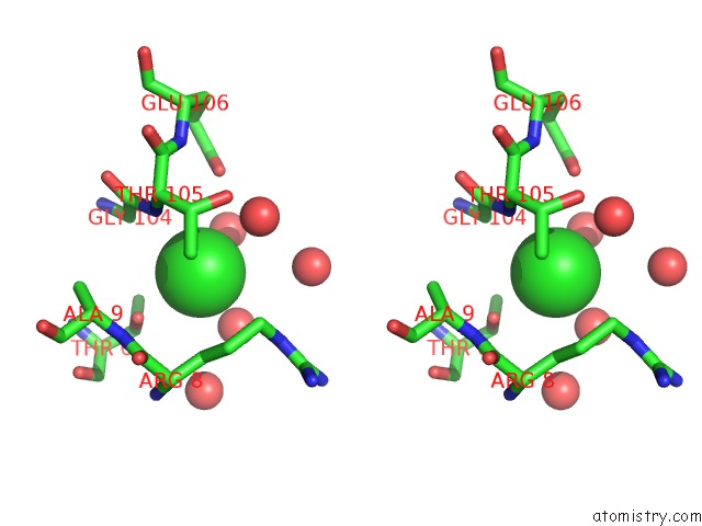
Chlorine binding site 2 out of 4 in 6cn8
Go back to
Chlorine binding site 2 out
of 4 in the High-Resolution Structure of CLPC1-Ntd Binding to Rufomycin-I

Mono view
Stereo pair view
Mono view
Stereo pair view
A full contact list of Chlorine with other atoms in the Cl binding
site number 2 of High-Resolution Structure of CLPC1-Ntd Binding to Rufomycin-I within 5.0Å range:
|
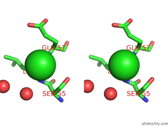
Chlorine binding site 3 out of 4 in 6cn8
Go back to
Chlorine binding site 3 out
of 4 in the High-Resolution Structure of CLPC1-Ntd Binding to Rufomycin-I

Mono view
Stereo pair view
Mono view
Stereo pair view
A full contact list of Chlorine with other atoms in the Cl binding
site number 3 of High-Resolution Structure of CLPC1-Ntd Binding to Rufomycin-I within 5.0Å range:
|
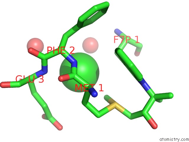
Chlorine binding site 4 out of 4 in 6cn8
Go back to
Chlorine binding site 4 out
of 4 in the High-Resolution Structure of CLPC1-Ntd Binding to Rufomycin-I
Mono view
Stereo pair view
Mono view
Stereo pair view
A full contact list of Chlorine with other atoms in the Cl binding
site number 4 of High-Resolution Structure of CLPC1-Ntd Binding to Rufomycin-I within 5.0Å range:
|
Reference:
N.M.Wolf,
H.Lee,
M.P.Choules,
G.F.Pauli,
R.Phansalkar,
J.R.Anderson,
W.Gao,
J.Ren,
B.D.Santarsiero,
H.Lee,
J.Cheng,
Y.Y.Jin,
N.A.Ho,
N.M.Duc,
J.W.Suh,
C.Abad-Zapatero,
S.Cho.
High-Resolution Structure of CLPC1-Rufomycin and Ligand Binding Studies Provide A Framework to Design and Optimize Anti-Tuberculosis Leads. Acs Infect Dis. V. 5 829 2019.
ISSN: ESSN 2373-8227
PubMed: 30990022
DOI: 10.1021/ACSINFECDIS.8B00276
Page generated: Sat Jul 12 12:28:39 2025
ISSN: ESSN 2373-8227
PubMed: 30990022
DOI: 10.1021/ACSINFECDIS.8B00276
Last articles
Fe in 2YXOFe in 2YRS
Fe in 2YXC
Fe in 2YNM
Fe in 2YVJ
Fe in 2YP1
Fe in 2YU2
Fe in 2YU1
Fe in 2YQB
Fe in 2YOO