Chlorine »
PDB 6cub-6d3h »
6d1q »
Chlorine in PDB 6d1q: Crystal Structure of E. Coli Rpph-Dapf Complex, Monomer
Enzymatic activity of Crystal Structure of E. Coli Rpph-Dapf Complex, Monomer
All present enzymatic activity of Crystal Structure of E. Coli Rpph-Dapf Complex, Monomer:
5.1.1.7;
5.1.1.7;
Protein crystallography data
The structure of Crystal Structure of E. Coli Rpph-Dapf Complex, Monomer, PDB code: 6d1q
was solved by
A.Gao,
A.Serganov,
with X-Ray Crystallography technique. A brief refinement statistics is given in the table below:
| Resolution Low / High (Å) | 123.65 / 2.15 |
| Space group | C 2 2 21 |
| Cell size a, b, c (Å), α, β, γ (°) | 161.440, 192.320, 50.719, 90.00, 90.00, 90.00 |
| R / Rfree (%) | 21.3 / 23.4 |
Chlorine Binding Sites:
The binding sites of Chlorine atom in the Crystal Structure of E. Coli Rpph-Dapf Complex, Monomer
(pdb code 6d1q). This binding sites where shown within
5.0 Angstroms radius around Chlorine atom.
In total 2 binding sites of Chlorine where determined in the Crystal Structure of E. Coli Rpph-Dapf Complex, Monomer, PDB code: 6d1q:
Jump to Chlorine binding site number: 1; 2;
In total 2 binding sites of Chlorine where determined in the Crystal Structure of E. Coli Rpph-Dapf Complex, Monomer, PDB code: 6d1q:
Jump to Chlorine binding site number: 1; 2;
Chlorine binding site 1 out of 2 in 6d1q
Go back to
Chlorine binding site 1 out
of 2 in the Crystal Structure of E. Coli Rpph-Dapf Complex, Monomer
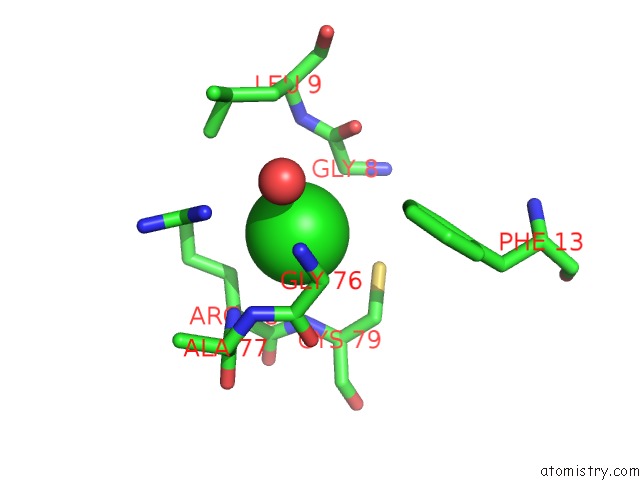
Mono view
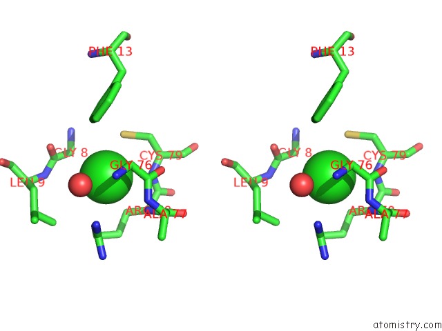
Stereo pair view
Mono view
Stereo pair view
A full contact list of Chlorine with other atoms in the Cl binding
site number 1 of Crystal Structure of E. Coli Rpph-Dapf Complex, Monomer within 5.0Å range:
|
Chlorine binding site 2 out of 2 in 6d1q
Go back to
Chlorine binding site 2 out
of 2 in the Crystal Structure of E. Coli Rpph-Dapf Complex, Monomer
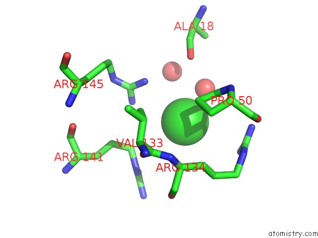
Mono view
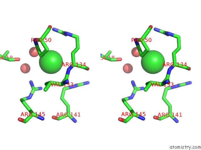
Stereo pair view
Mono view
Stereo pair view
A full contact list of Chlorine with other atoms in the Cl binding
site number 2 of Crystal Structure of E. Coli Rpph-Dapf Complex, Monomer within 5.0Å range:
|
Reference:
A.Gao,
N.Vasilyev,
D.J.Luciano,
R.Levenson-Palmer,
J.Richards,
W.M.Marsiglia,
N.J.Traaseth,
J.G.Belasco,
A.Serganov.
Structural and Kinetic Insights Into Stimulation of Rpph-Dependent Rna Degradation By the Metabolic Enzyme Dapf. Nucleic Acids Res. V. 46 6841 2018.
ISSN: ESSN 1362-4962
PubMed: 29733359
DOI: 10.1093/NAR/GKY327
Page generated: Sat Jul 27 21:15:37 2024
ISSN: ESSN 1362-4962
PubMed: 29733359
DOI: 10.1093/NAR/GKY327
Last articles
Zn in 9J0NZn in 9J0O
Zn in 9J0P
Zn in 9FJX
Zn in 9EKB
Zn in 9C0F
Zn in 9CAH
Zn in 9CH0
Zn in 9CH3
Zn in 9CH1