Chlorine »
PDB 6d3i-6ddg »
6d4c »
Chlorine in PDB 6d4c: Crystal Structure of Candida Boidinii Formate Dehydrogenase V123G Mutant Complexed with Nad+ and Azide
Enzymatic activity of Crystal Structure of Candida Boidinii Formate Dehydrogenase V123G Mutant Complexed with Nad+ and Azide
All present enzymatic activity of Crystal Structure of Candida Boidinii Formate Dehydrogenase V123G Mutant Complexed with Nad+ and Azide:
1.17.1.9;
1.17.1.9;
Protein crystallography data
The structure of Crystal Structure of Candida Boidinii Formate Dehydrogenase V123G Mutant Complexed with Nad+ and Azide, PDB code: 6d4c
was solved by
Q.Guo,
H.Ye,
L.Gakhar,
C.M.Cheatum,
A.Kohen,
with X-Ray Crystallography technique. A brief refinement statistics is given in the table below:
| Resolution Low / High (Å) | 45.99 / 1.45 |
| Space group | P 1 21 1 |
| Cell size a, b, c (Å), α, β, γ (°) | 51.188, 115.899, 65.469, 90.00, 108.56, 90.00 |
| R / Rfree (%) | 15.9 / 18.5 |
Chlorine Binding Sites:
The binding sites of Chlorine atom in the Crystal Structure of Candida Boidinii Formate Dehydrogenase V123G Mutant Complexed with Nad+ and Azide
(pdb code 6d4c). This binding sites where shown within
5.0 Angstroms radius around Chlorine atom.
In total 2 binding sites of Chlorine where determined in the Crystal Structure of Candida Boidinii Formate Dehydrogenase V123G Mutant Complexed with Nad+ and Azide, PDB code: 6d4c:
Jump to Chlorine binding site number: 1; 2;
In total 2 binding sites of Chlorine where determined in the Crystal Structure of Candida Boidinii Formate Dehydrogenase V123G Mutant Complexed with Nad+ and Azide, PDB code: 6d4c:
Jump to Chlorine binding site number: 1; 2;
Chlorine binding site 1 out of 2 in 6d4c
Go back to
Chlorine binding site 1 out
of 2 in the Crystal Structure of Candida Boidinii Formate Dehydrogenase V123G Mutant Complexed with Nad+ and Azide
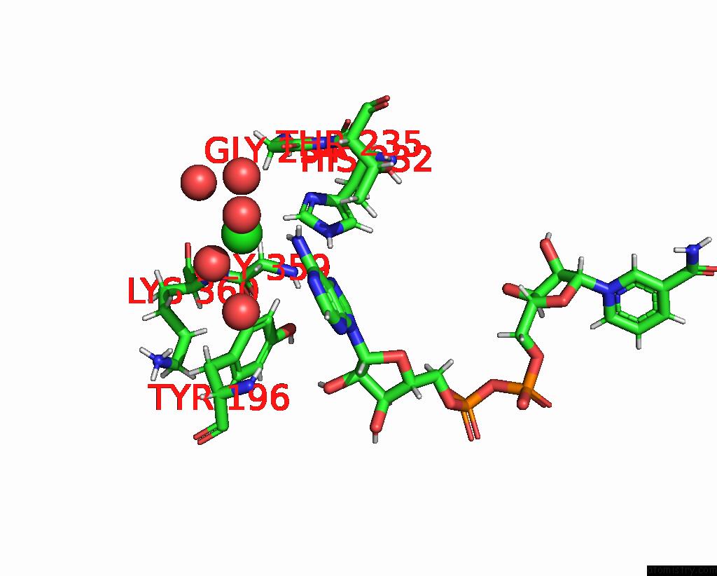
Mono view
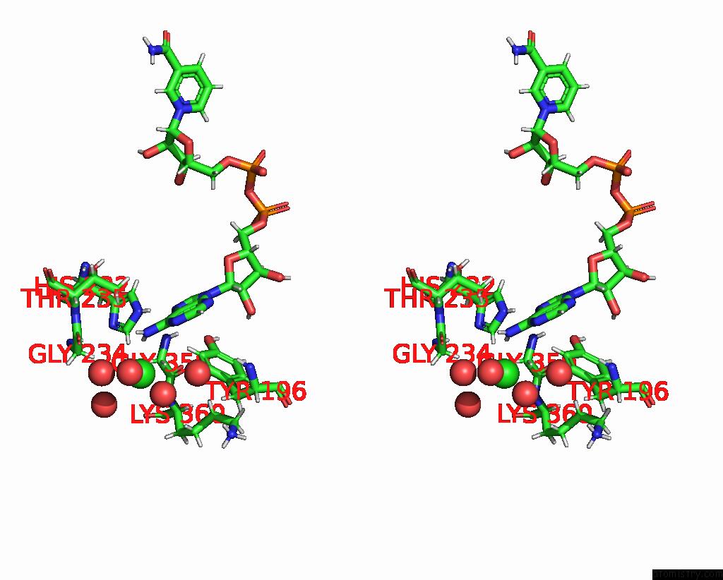
Stereo pair view
Mono view
Stereo pair view
A full contact list of Chlorine with other atoms in the Cl binding
site number 1 of Crystal Structure of Candida Boidinii Formate Dehydrogenase V123G Mutant Complexed with Nad+ and Azide within 5.0Å range:
|
Chlorine binding site 2 out of 2 in 6d4c
Go back to
Chlorine binding site 2 out
of 2 in the Crystal Structure of Candida Boidinii Formate Dehydrogenase V123G Mutant Complexed with Nad+ and Azide
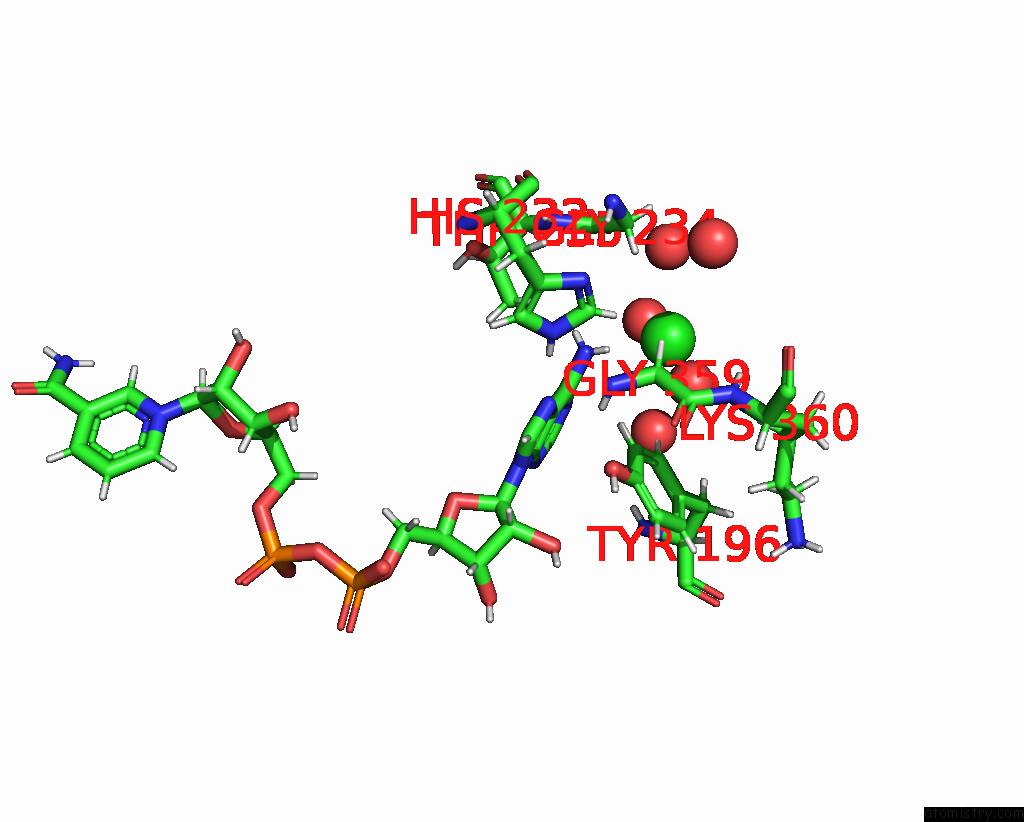
Mono view
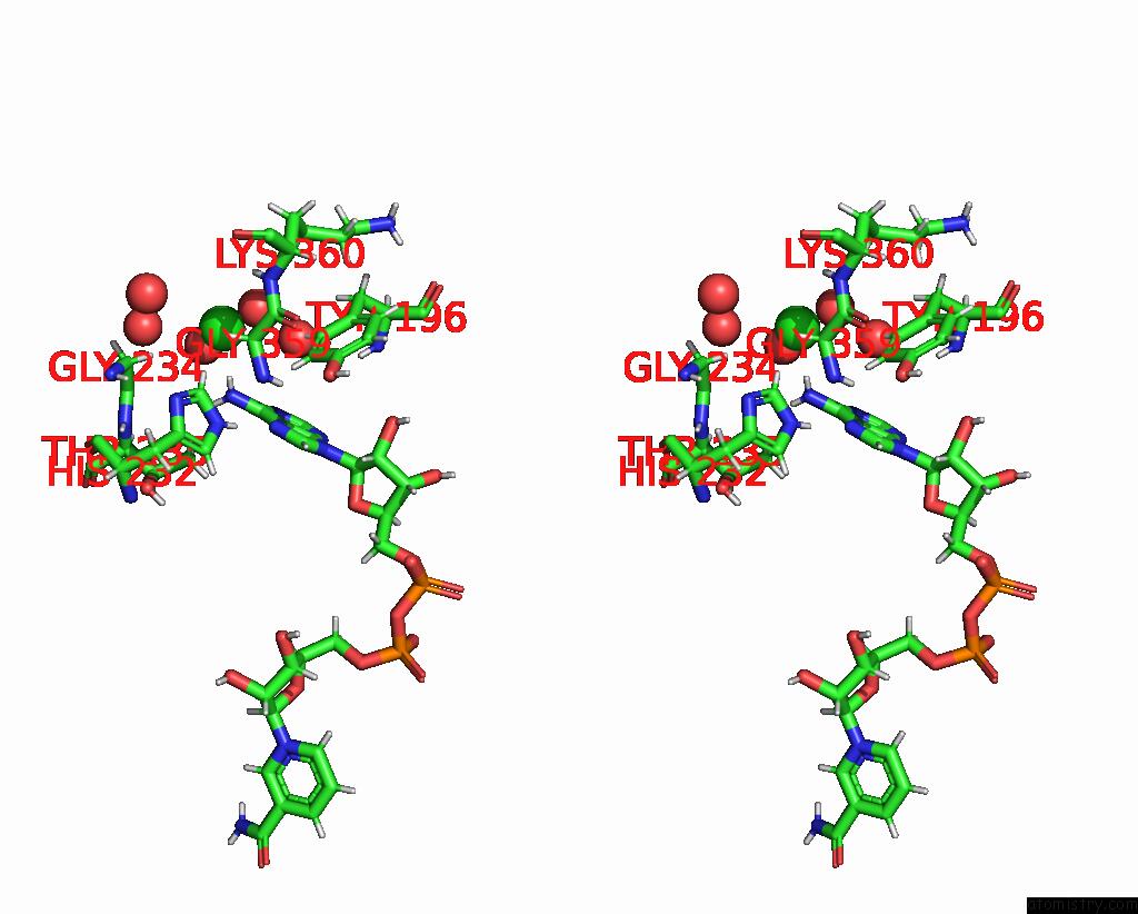
Stereo pair view
Mono view
Stereo pair view
A full contact list of Chlorine with other atoms in the Cl binding
site number 2 of Crystal Structure of Candida Boidinii Formate Dehydrogenase V123G Mutant Complexed with Nad+ and Azide within 5.0Å range:
|
Reference:
P.L.Pagano,
Q.Guo,
C.Ranasinghe,
E.Schroeder,
K.Robben,
F.Hase,
H.Ye,
K.Wickersham,
A.Aspuru-Guzik,
D.T.Major,
L.Gakhar,
A.Kohen,
C.M.Cheatum.
Oscillatory Active-Site Motions Correlate with Kinetic Isotope Effects in Formate Dehydrogenase Acs Catalysis 2019.
ISSN: ESSN 2155-5435
DOI: 10.1021/ACSCATAL.9B03345
Page generated: Sat Jul 12 12:43:13 2025
ISSN: ESSN 2155-5435
DOI: 10.1021/ACSCATAL.9B03345
Last articles
F in 4FQ4F in 4FPH
F in 4FNZ
F in 4FOD
F in 4FOR
F in 4FMD
F in 4FME
F in 4FMC
F in 4FMB
F in 4FM5