Chlorine »
PDB 6dow-6dy1 »
6dt1 »
Chlorine in PDB 6dt1: Crystal Structure of the Ligase From Bacteriophage T4 Complexed with Dna Intermediate
Enzymatic activity of Crystal Structure of the Ligase From Bacteriophage T4 Complexed with Dna Intermediate
All present enzymatic activity of Crystal Structure of the Ligase From Bacteriophage T4 Complexed with Dna Intermediate:
6.5.1.1;
6.5.1.1;
Protein crystallography data
The structure of Crystal Structure of the Ligase From Bacteriophage T4 Complexed with Dna Intermediate, PDB code: 6dt1
was solved by
K.Shi,
H.Aihara,
with X-Ray Crystallography technique. A brief refinement statistics is given in the table below:
| Resolution Low / High (Å) | 113.02 / 2.75 |
| Space group | P 1 |
| Cell size a, b, c (Å), α, β, γ (°) | 63.526, 67.429, 114.635, 88.78, 80.94, 62.97 |
| R / Rfree (%) | 17 / 21.9 |
Other elements in 6dt1:
The structure of Crystal Structure of the Ligase From Bacteriophage T4 Complexed with Dna Intermediate also contains other interesting chemical elements:
| Magnesium | (Mg) | 1 atom |
Chlorine Binding Sites:
The binding sites of Chlorine atom in the Crystal Structure of the Ligase From Bacteriophage T4 Complexed with Dna Intermediate
(pdb code 6dt1). This binding sites where shown within
5.0 Angstroms radius around Chlorine atom.
In total 2 binding sites of Chlorine where determined in the Crystal Structure of the Ligase From Bacteriophage T4 Complexed with Dna Intermediate, PDB code: 6dt1:
Jump to Chlorine binding site number: 1; 2;
In total 2 binding sites of Chlorine where determined in the Crystal Structure of the Ligase From Bacteriophage T4 Complexed with Dna Intermediate, PDB code: 6dt1:
Jump to Chlorine binding site number: 1; 2;
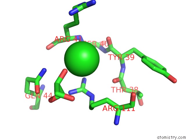
Chlorine binding site 1 out of 2 in 6dt1
Go back to
Chlorine binding site 1 out
of 2 in the Crystal Structure of the Ligase From Bacteriophage T4 Complexed with Dna Intermediate
Mono view
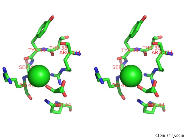
Stereo pair view
Mono view
Stereo pair view
A full contact list of Chlorine with other atoms in the Cl binding
site number 1 of Crystal Structure of the Ligase From Bacteriophage T4 Complexed with Dna Intermediate within 5.0Å range:
|
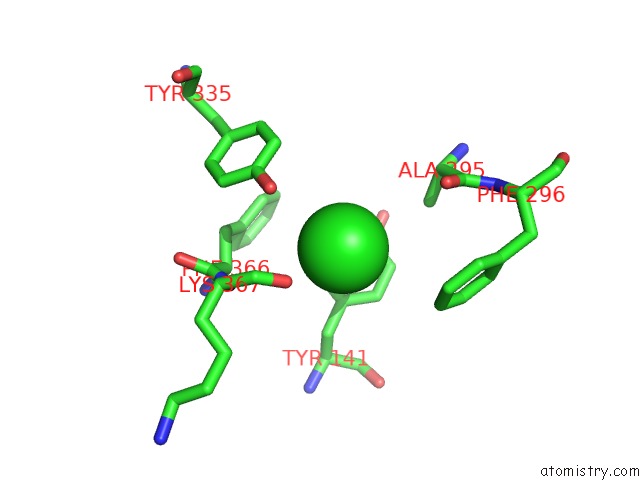
Chlorine binding site 2 out of 2 in 6dt1
Go back to
Chlorine binding site 2 out
of 2 in the Crystal Structure of the Ligase From Bacteriophage T4 Complexed with Dna Intermediate
Mono view
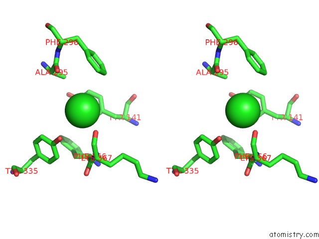
Stereo pair view
Mono view
Stereo pair view
A full contact list of Chlorine with other atoms in the Cl binding
site number 2 of Crystal Structure of the Ligase From Bacteriophage T4 Complexed with Dna Intermediate within 5.0Å range:
|
Reference:
K.Shi,
T.E.Bohl,
J.Park,
A.Zasada,
S.Malik,
S.Banerjee,
V.Tran,
N.Li,
Z.Yin,
F.Kurniawan,
K.Orellana,
H.Aihara.
T4 Dna Ligase Structure Reveals A Prototypical Atp-Dependent Ligase with A Unique Mode of Sliding Clamp Interaction. Nucleic Acids Res. V. 46 10474 2018.
ISSN: ESSN 1362-4962
PubMed: 30169742
DOI: 10.1093/NAR/GKY776
Page generated: Sat Jul 27 21:48:43 2024
ISSN: ESSN 1362-4962
PubMed: 30169742
DOI: 10.1093/NAR/GKY776
Last articles
Zn in 9J0NZn in 9J0O
Zn in 9J0P
Zn in 9FJX
Zn in 9EKB
Zn in 9C0F
Zn in 9CAH
Zn in 9CH0
Zn in 9CH3
Zn in 9CH1