Chlorine in PDB 6eg5: The Structure of Sb-1-202-Tubulin Complex
Protein crystallography data
The structure of The Structure of Sb-1-202-Tubulin Complex, PDB code: 6eg5
was solved by
A.K.Banerjee,
Y.Wang,
H.Chen,
D.Miller,
W.Li,
with X-Ray Crystallography technique. A brief refinement statistics is given in the table below:
Resolution Low / High (Å)
|
50.00 /
2.45
|
Space group
|
P 21 21 21
|
Cell size a, b, c (Å), α, β, γ (°)
|
105.541,
158.033,
181.870,
90.00,
90.00,
90.00
|
R / Rfree (%)
|
19.6 /
23.6
|
Other elements in 6eg5:
The structure of The Structure of Sb-1-202-Tubulin Complex also contains other interesting chemical elements:
Chlorine Binding Sites:
The binding sites of Chlorine atom in the The Structure of Sb-1-202-Tubulin Complex
(pdb code 6eg5). This binding sites where shown within
5.0 Angstroms radius around Chlorine atom.
In total 3 binding sites of Chlorine where determined in the
The Structure of Sb-1-202-Tubulin Complex, PDB code: 6eg5:
Jump to Chlorine binding site number:
1;
2;
3;
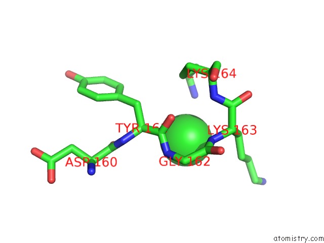
Chlorine binding site 1 out
of 3 in 6eg5
Go back to
Chlorine Binding Sites List in 6eg5
Chlorine binding site 1 out
of 3 in the The Structure of Sb-1-202-Tubulin Complex
 Mono view
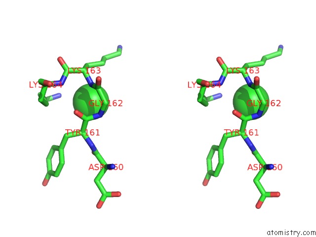
 Stereo pair view
|
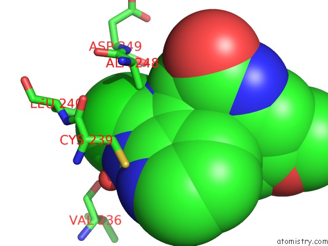
A full contact list of Chlorine with other atoms in the Cl binding
site number 1 of The Structure of Sb-1-202-Tubulin Complex within 5.0Å range:
probe
|
atom
|
residue
|
distance (Å)
|
B
|
Occ
|
A:Cl504
b:74.2
occ:1.00
|
N
|
A:LYS163
|
3.2
|
61.4
|
1.0
|
NZ
|
A:LYS164
|
3.4
|
65.1
|
1.0
|
N
|
A:GLY162
|
3.4
|
45.3
|
1.0
|
C
|
A:TYR161
|
3.5
|
44.2
|
1.0
|
CB
|
A:LYS163
|
3.6
|
72.0
|
1.0
|
CA
|
A:TYR161
|
3.7
|
45.5
|
1.0
|
CG
|
A:LYS163
|
3.9
|
77.1
|
1.0
|
CA
|
A:LYS163
|
3.9
|
66.6
|
1.0
|
O
|
A:TYR161
|
4.0
|
47.7
|
1.0
|
C
|
A:GLY162
|
4.1
|
52.9
|
1.0
|
CA
|
A:GLY162
|
4.2
|
49.4
|
1.0
|
CD
|
A:LYS163
|
4.4
|
82.7
|
1.0
|
CG
|
A:LYS164
|
4.5
|
56.1
|
1.0
|
CE
|
A:LYS164
|
4.5
|
62.0
|
1.0
|
N
|
A:LYS164
|
4.6
|
57.4
|
1.0
|
CB
|
A:TYR161
|
4.6
|
43.6
|
1.0
|
C
|
A:LYS163
|
4.6
|
63.4
|
1.0
|
N
|
A:TYR161
|
4.8
|
49.4
|
1.0
|
O
|
A:ASP160
|
4.8
|
49.5
|
1.0
|
CD
|
A:LYS164
|
4.9
|
60.0
|
1.0
|
|
Chlorine binding site 2 out
of 3 in 6eg5
Go back to
Chlorine Binding Sites List in 6eg5
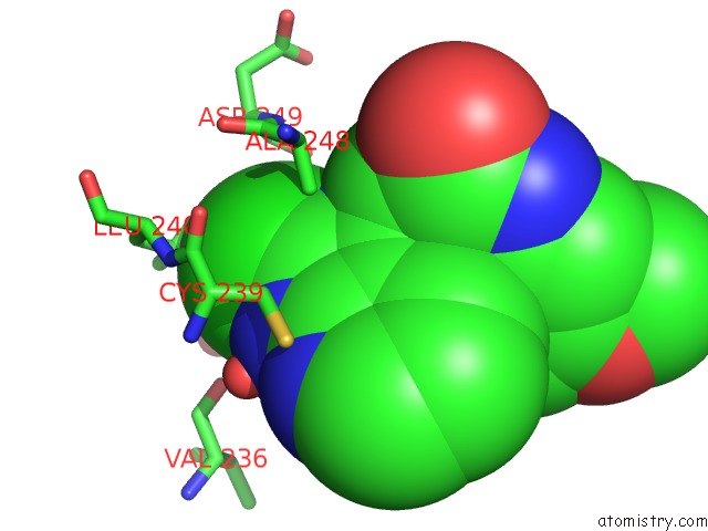
Chlorine binding site 2 out
of 3 in the The Structure of Sb-1-202-Tubulin Complex
 Mono view
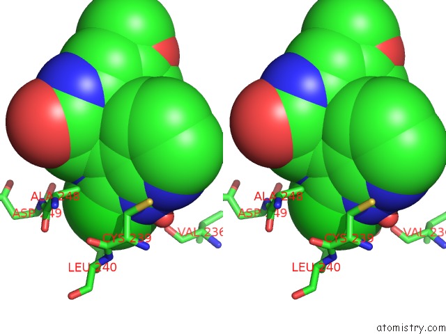
 Stereo pair view
|
A full contact list of Chlorine with other atoms in the Cl binding
site number 2 of The Structure of Sb-1-202-Tubulin Complex within 5.0Å range:
probe
|
atom
|
residue
|
distance (Å)
|
B
|
Occ
|
B:Cl506
b:39.6
occ:1.00
|
CL
|
B:J7S506
|
0.0
|
39.6
|
1.0
|
C6
|
B:J7S506
|
1.7
|
33.0
|
1.0
|
N1
|
B:J7S506
|
2.6
|
32.3
|
1.0
|
N2
|
B:J7S506
|
2.6
|
33.1
|
1.0
|
CD2
|
B:LEU240
|
3.3
|
30.8
|
1.0
|
CB
|
B:ALA248
|
3.6
|
41.3
|
1.0
|
CG
|
B:LEU240
|
3.7
|
32.0
|
1.0
|
C5
|
B:J7S506
|
3.8
|
29.1
|
1.0
|
C7
|
B:J7S506
|
3.9
|
31.9
|
1.0
|
O
|
B:HOH692
|
3.9
|
40.9
|
1.0
|
CD2
|
B:LEU253
|
4.1
|
25.9
|
1.0
|
CB
|
B:LEU253
|
4.2
|
25.1
|
1.0
|
CB
|
B:CYS239
|
4.3
|
34.7
|
1.0
|
O
|
B:VAL236
|
4.4
|
27.9
|
1.0
|
CD1
|
B:LEU253
|
4.4
|
25.6
|
1.0
|
CD1
|
B:LEU240
|
4.4
|
32.5
|
1.0
|
C8
|
B:J7S506
|
4.4
|
30.8
|
1.0
|
CG
|
B:LEU253
|
4.4
|
25.5
|
1.0
|
O
|
B:HOH607
|
4.6
|
29.7
|
1.0
|
O
|
B:ASP249
|
4.6
|
28.1
|
1.0
|
N
|
B:LEU240
|
4.6
|
35.2
|
1.0
|
C
|
B:CYS239
|
4.7
|
36.2
|
1.0
|
N
|
B:J7S506
|
4.9
|
26.8
|
1.0
|
CA
|
B:ALA248
|
4.9
|
42.0
|
1.0
|
N
|
B:ASP249
|
4.9
|
34.1
|
1.0
|
CB
|
B:LEU240
|
4.9
|
33.0
|
1.0
|
C
|
B:ALA248
|
4.9
|
35.7
|
1.0
|
N3
|
B:J7S506
|
5.0
|
31.7
|
1.0
|
C
|
B:ASP249
|
5.0
|
29.1
|
1.0
|
|
Chlorine binding site 3 out
of 3 in 6eg5
Go back to
Chlorine Binding Sites List in 6eg5
Chlorine binding site 3 out
of 3 in the The Structure of Sb-1-202-Tubulin Complex
 Mono view
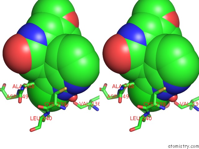
 Stereo pair view
|
A full contact list of Chlorine with other atoms in the Cl binding
site number 3 of The Structure of Sb-1-202-Tubulin Complex within 5.0Å range:
probe
|
atom
|
residue
|
distance (Å)
|
B
|
Occ
|
D:Cl603
b:48.8
occ:1.00
|
CL
|
D:J7S603
|
0.0
|
48.8
|
1.0
|
C6
|
D:J7S603
|
1.8
|
45.6
|
1.0
|
N1
|
D:J7S603
|
2.6
|
42.9
|
1.0
|
N2
|
D:J7S603
|
2.7
|
44.5
|
1.0
|
CD2
|
D:LEU240
|
3.4
|
38.0
|
1.0
|
CB
|
D:ALA248
|
3.6
|
44.1
|
1.0
|
CG
|
D:LEU240
|
3.7
|
38.3
|
1.0
|
C5
|
D:J7S603
|
3.9
|
38.0
|
1.0
|
CB
|
D:LEU253
|
3.9
|
30.2
|
1.0
|
C7
|
D:J7S603
|
4.0
|
42.2
|
1.0
|
CD2
|
D:LEU253
|
4.1
|
32.6
|
1.0
|
O
|
D:HOH734
|
4.1
|
60.9
|
1.0
|
CD1
|
D:LEU253
|
4.2
|
31.4
|
1.0
|
CG
|
D:LEU253
|
4.3
|
31.4
|
1.0
|
O
|
D:ASP249
|
4.3
|
28.9
|
1.0
|
CB
|
D:CYS239
|
4.3
|
44.0
|
1.0
|
N
|
D:LEU240
|
4.4
|
40.2
|
1.0
|
O
|
D:VAL236
|
4.5
|
47.1
|
1.0
|
C8
|
D:J7S603
|
4.5
|
41.0
|
1.0
|
CD1
|
D:LEU240
|
4.6
|
39.7
|
1.0
|
C
|
D:CYS239
|
4.6
|
42.2
|
1.0
|
N
|
D:ASP249
|
4.7
|
34.0
|
1.0
|
O
|
D:HOH709
|
4.7
|
47.4
|
1.0
|
C
|
D:ASP249
|
4.8
|
31.4
|
1.0
|
C
|
D:ALA248
|
4.8
|
38.0
|
1.0
|
CA
|
D:ALA248
|
4.9
|
41.6
|
1.0
|
CB
|
D:LEU240
|
4.9
|
37.7
|
1.0
|
N
|
D:J7S603
|
4.9
|
33.2
|
1.0
|
CA
|
D:LEU253
|
4.9
|
28.6
|
1.0
|
CA
|
D:CYS239
|
4.9
|
42.7
|
1.0
|
CA
|
D:LEU240
|
4.9
|
39.6
|
1.0
|
|
Reference:
H.Chen,
Y.Wang,
W.Li.
The Structure of Sb-1-202/Sb-2-204-Tubulin Complex To Be Published.
Page generated: Sat Jul 27 22:21:41 2024
|