Chlorine »
PDB 6el4-6erc »
6eo9 »
Chlorine in PDB 6eo9: Crystal Structure of Thrombin in Complex with A Novel Glucose- Conjugated Potent Inhibitor
Enzymatic activity of Crystal Structure of Thrombin in Complex with A Novel Glucose- Conjugated Potent Inhibitor
All present enzymatic activity of Crystal Structure of Thrombin in Complex with A Novel Glucose- Conjugated Potent Inhibitor:
3.4.21.5;
3.4.21.5;
Protein crystallography data
The structure of Crystal Structure of Thrombin in Complex with A Novel Glucose- Conjugated Potent Inhibitor, PDB code: 6eo9
was solved by
B.D.Belviso,
R.Caliandro,
B.M.Aresta,
M.De Candia,
C.D.Altomare,
with X-Ray Crystallography technique. A brief refinement statistics is given in the table below:
| Resolution Low / High (Å) | 37.86 / 1.84 |
| Space group | C 1 2 1 |
| Cell size a, b, c (Å), α, β, γ (°) | 67.460, 71.640, 71.800, 90.00, 100.21, 90.00 |
| R / Rfree (%) | 19.8 / 24.8 |
Chlorine Binding Sites:
The binding sites of Chlorine atom in the Crystal Structure of Thrombin in Complex with A Novel Glucose- Conjugated Potent Inhibitor
(pdb code 6eo9). This binding sites where shown within
5.0 Angstroms radius around Chlorine atom.
In total only one binding site of Chlorine was determined in the Crystal Structure of Thrombin in Complex with A Novel Glucose- Conjugated Potent Inhibitor, PDB code: 6eo9:
In total only one binding site of Chlorine was determined in the Crystal Structure of Thrombin in Complex with A Novel Glucose- Conjugated Potent Inhibitor, PDB code: 6eo9:
Chlorine binding site 1 out of 1 in 6eo9
Go back to
Chlorine binding site 1 out
of 1 in the Crystal Structure of Thrombin in Complex with A Novel Glucose- Conjugated Potent Inhibitor
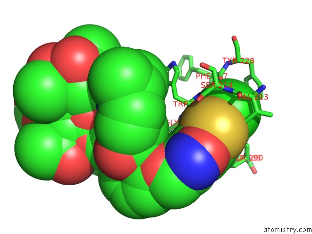
Mono view
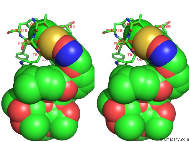
Stereo pair view
Mono view
Stereo pair view
A full contact list of Chlorine with other atoms in the Cl binding
site number 1 of Crystal Structure of Thrombin in Complex with A Novel Glucose- Conjugated Potent Inhibitor within 5.0Å range:
|
Reference:
B.D.Belviso,
R.Caliandro,
M.De Candia,
G.Zaetta,
G.Lopopolo,
F.Incampo,
M.Colucci,
C.D.Altomare.
How A Beta-D-Glucoside Side Chain Enhances Binding Affinity to Thrombin of Inhibitors Bearing 2-Chlorothiophene As P1 Moiety: Crystallography, Fragment Deconstruction Study, and Evaluation of Antithrombotic Properties. J. Med. Chem. V. 57 8563 2014.
ISSN: ISSN 1520-4804
PubMed: 25268757
DOI: 10.1021/JM5010754
Page generated: Sat Jul 27 22:30:10 2024
ISSN: ISSN 1520-4804
PubMed: 25268757
DOI: 10.1021/JM5010754
Last articles
Zn in 9J0NZn in 9J0O
Zn in 9J0P
Zn in 9FJX
Zn in 9EKB
Zn in 9C0F
Zn in 9CAH
Zn in 9CH0
Zn in 9CH3
Zn in 9CH1