Chlorine »
PDB 6erd-6ezo »
6ero »
Chlorine in PDB 6ero: Structure of Human TFB2M
Protein crystallography data
The structure of Structure of Human TFB2M, PDB code: 6ero
was solved by
H.S.Hillen,
Y.I.Morozov,
A.Sarfallah,
D.Temiakov,
P.Cramer,
with X-Ray Crystallography technique. A brief refinement statistics is given in the table below:
| Resolution Low / High (Å) | 43.53 / 1.75 |
| Space group | P 1 21 1 |
| Cell size a, b, c (Å), α, β, γ (°) | 43.950, 165.650, 44.730, 90.00, 97.97, 90.00 |
| R / Rfree (%) | 19.2 / 21.4 |
Chlorine Binding Sites:
The binding sites of Chlorine atom in the Structure of Human TFB2M
(pdb code 6ero). This binding sites where shown within
5.0 Angstroms radius around Chlorine atom.
In total 2 binding sites of Chlorine where determined in the Structure of Human TFB2M, PDB code: 6ero:
Jump to Chlorine binding site number: 1; 2;
In total 2 binding sites of Chlorine where determined in the Structure of Human TFB2M, PDB code: 6ero:
Jump to Chlorine binding site number: 1; 2;
Chlorine binding site 1 out of 2 in 6ero
Go back to
Chlorine binding site 1 out
of 2 in the Structure of Human TFB2M
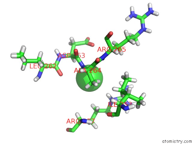
Mono view
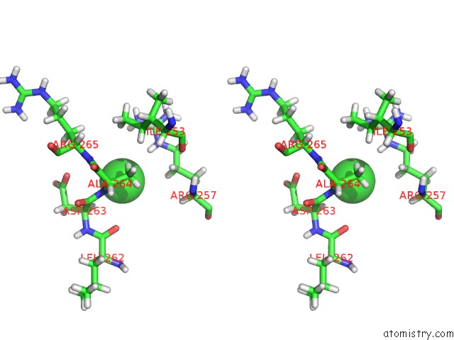
Stereo pair view
Mono view
Stereo pair view
A full contact list of Chlorine with other atoms in the Cl binding
site number 1 of Structure of Human TFB2M within 5.0Å range:
|
Chlorine binding site 2 out of 2 in 6ero
Go back to
Chlorine binding site 2 out
of 2 in the Structure of Human TFB2M
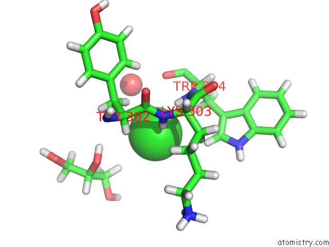
Mono view
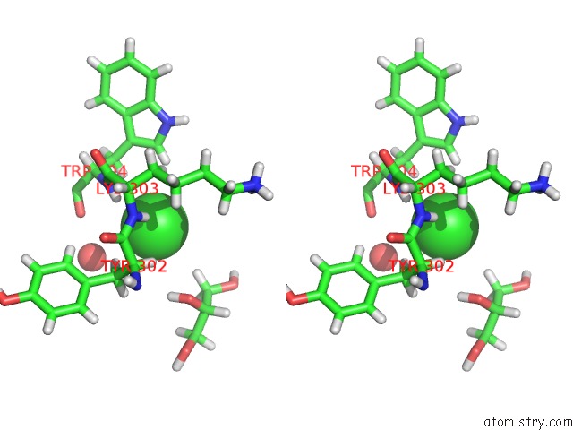
Stereo pair view
Mono view
Stereo pair view
A full contact list of Chlorine with other atoms in the Cl binding
site number 2 of Structure of Human TFB2M within 5.0Å range:
|
Reference:
H.S.Hillen,
Y.I.Morozov,
A.Sarfallah,
D.Temiakov,
P.Cramer.
Structural Basis of Mitochondrial Transcription Initiation. Cell V. 171 1072 2017.
ISSN: ISSN 1097-4172
PubMed: 29149603
DOI: 10.1016/J.CELL.2017.10.036
Page generated: Sat Jul 27 22:39:03 2024
ISSN: ISSN 1097-4172
PubMed: 29149603
DOI: 10.1016/J.CELL.2017.10.036
Last articles
Zn in 9J0NZn in 9J0O
Zn in 9J0P
Zn in 9FJX
Zn in 9EKB
Zn in 9C0F
Zn in 9CAH
Zn in 9CH0
Zn in 9CH3
Zn in 9CH1