Chlorine »
PDB 6erd-6ezo »
6eup »
Chlorine in PDB 6eup: Crystal Structure of Neisseria Meningitidis Nada Variant 3 Double Mutant A33I-I38L
Protein crystallography data
The structure of Crystal Structure of Neisseria Meningitidis Nada Variant 3 Double Mutant A33I-I38L, PDB code: 6eup
was solved by
L.Dello Iacono,
A.Liguori,
E.Malito,
M.J.Bottomley,
with X-Ray Crystallography technique. A brief refinement statistics is given in the table below:
| Resolution Low / High (Å) | 63.65 / 2.65 |
| Space group | C 1 2 1 |
| Cell size a, b, c (Å), α, β, γ (°) | 68.919, 39.750, 255.630, 90.00, 95.13, 90.00 |
| R / Rfree (%) | 19.8 / 23.6 |
Chlorine Binding Sites:
The binding sites of Chlorine atom in the Crystal Structure of Neisseria Meningitidis Nada Variant 3 Double Mutant A33I-I38L
(pdb code 6eup). This binding sites where shown within
5.0 Angstroms radius around Chlorine atom.
In total only one binding site of Chlorine was determined in the Crystal Structure of Neisseria Meningitidis Nada Variant 3 Double Mutant A33I-I38L, PDB code: 6eup:
In total only one binding site of Chlorine was determined in the Crystal Structure of Neisseria Meningitidis Nada Variant 3 Double Mutant A33I-I38L, PDB code: 6eup:
Chlorine binding site 1 out of 1 in 6eup
Go back to
Chlorine binding site 1 out
of 1 in the Crystal Structure of Neisseria Meningitidis Nada Variant 3 Double Mutant A33I-I38L
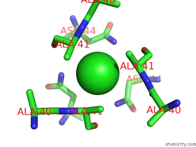
Mono view
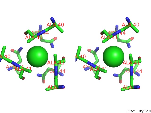
Stereo pair view
Mono view
Stereo pair view
A full contact list of Chlorine with other atoms in the Cl binding
site number 1 of Crystal Structure of Neisseria Meningitidis Nada Variant 3 Double Mutant A33I-I38L within 5.0Å range:
|
Reference:
A.Liguori,
L.Dello Iacono,
G.Maruggi,
B.Benucci,
M.Merola,
P.Lo Surdo,
J.Lopez-Sagaseta,
M.Pizza,
E.Malito,
M.J.Bottomley.
NADA3 Structures Reveal Undecad Coiled Coils and LOX1 Binding Regions Competed By Meningococcus B Vaccine-Elicited Human Antibodies. Mbio V. 9 2018.
ISSN: ESSN 2150-7511
PubMed: 30327444
DOI: 10.1128/MBIO.01914-18
Page generated: Sat Jul 27 22:41:28 2024
ISSN: ESSN 2150-7511
PubMed: 30327444
DOI: 10.1128/MBIO.01914-18
Last articles
Zn in 9J0NZn in 9J0O
Zn in 9J0P
Zn in 9FJX
Zn in 9EKB
Zn in 9C0F
Zn in 9CAH
Zn in 9CH0
Zn in 9CH3
Zn in 9CH1