Chlorine »
PDB 6ezp-6f4d »
6f04 »
Chlorine in PDB 6f04: N-Acetylglucosamine-2-Epimerase
Protein crystallography data
The structure of N-Acetylglucosamine-2-Epimerase, PDB code: 6f04
was solved by
M.J.Halsoer,
U.Rothweiler,
with X-Ray Crystallography technique. A brief refinement statistics is given in the table below:
| Resolution Low / High (Å) | 48.88 / 1.70 |
| Space group | P 42 21 2 |
| Cell size a, b, c (Å), α, β, γ (°) | 142.423, 142.423, 52.046, 90.00, 90.00, 90.00 |
| R / Rfree (%) | 17.9 / 20.8 |
Chlorine Binding Sites:
The binding sites of Chlorine atom in the N-Acetylglucosamine-2-Epimerase
(pdb code 6f04). This binding sites where shown within
5.0 Angstroms radius around Chlorine atom.
In total only one binding site of Chlorine was determined in the N-Acetylglucosamine-2-Epimerase, PDB code: 6f04:
In total only one binding site of Chlorine was determined in the N-Acetylglucosamine-2-Epimerase, PDB code: 6f04:
Chlorine binding site 1 out of 1 in 6f04
Go back to
Chlorine binding site 1 out
of 1 in the N-Acetylglucosamine-2-Epimerase

Mono view
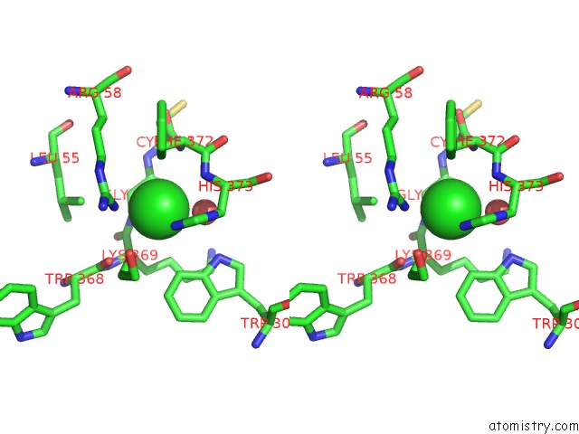
Stereo pair view
Mono view
Stereo pair view
A full contact list of Chlorine with other atoms in the Cl binding
site number 1 of N-Acetylglucosamine-2-Epimerase within 5.0Å range:
|
Reference:
M.J.H.Halsor,
U.Rothweiler,
B.Altermark,
I.L.U.Raeder.
The Crystal Structure of the N-Acetylglucosamine 2-Epimerase From Nostoc Sp. KVJ10 Reveals the True Dimer. Acta Crystallogr D Struct V. 75 90 2019BIOL.
ISSN: ISSN 2059-7983
PubMed: 30644848
DOI: 10.1107/S2059798318017047
Page generated: Sat Jul 27 22:45:43 2024
ISSN: ISSN 2059-7983
PubMed: 30644848
DOI: 10.1107/S2059798318017047
Last articles
Zn in 9J0NZn in 9J0O
Zn in 9J0P
Zn in 9FJX
Zn in 9EKB
Zn in 9C0F
Zn in 9CAH
Zn in 9CH0
Zn in 9CH3
Zn in 9CH1