Chlorine »
PDB 6fbv-6fkv »
6ffh »
Chlorine in PDB 6ffh: Crystal Structure of MGLUR5 in Complex with Fenobam at 2.65 A
Enzymatic activity of Crystal Structure of MGLUR5 in Complex with Fenobam at 2.65 A
All present enzymatic activity of Crystal Structure of MGLUR5 in Complex with Fenobam at 2.65 A:
3.2.1.17;
3.2.1.17;
Protein crystallography data
The structure of Crystal Structure of MGLUR5 in Complex with Fenobam at 2.65 A, PDB code: 6ffh
was solved by
J.A.Christopher,
Z.Orgovan,
M.Congreve,
A.S.Dore,
J.C.Errey,
F.H.Marshall,
J.S.Mason,
K.Okrasa,
P.Rucktooa,
M.J.Serrano-Vega,
G.G.Ferenczy,
G.M.Keseru,
with X-Ray Crystallography technique. A brief refinement statistics is given in the table below:
| Resolution Low / High (Å) | 34.50 / 2.65 |
| Space group | C 1 2 1 |
| Cell size a, b, c (Å), α, β, γ (°) | 143.217, 43.478, 82.383, 90.00, 99.22, 90.00 |
| R / Rfree (%) | 24.4 / 26.7 |
Chlorine Binding Sites:
The binding sites of Chlorine atom in the Crystal Structure of MGLUR5 in Complex with Fenobam at 2.65 A
(pdb code 6ffh). This binding sites where shown within
5.0 Angstroms radius around Chlorine atom.
In total only one binding site of Chlorine was determined in the Crystal Structure of MGLUR5 in Complex with Fenobam at 2.65 A, PDB code: 6ffh:
In total only one binding site of Chlorine was determined in the Crystal Structure of MGLUR5 in Complex with Fenobam at 2.65 A, PDB code: 6ffh:
Chlorine binding site 1 out of 1 in 6ffh
Go back to
Chlorine binding site 1 out
of 1 in the Crystal Structure of MGLUR5 in Complex with Fenobam at 2.65 A
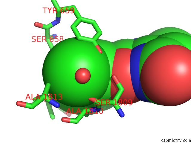
Mono view
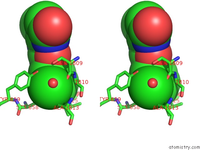
Stereo pair view
Mono view
Stereo pair view
A full contact list of Chlorine with other atoms in the Cl binding
site number 1 of Crystal Structure of MGLUR5 in Complex with Fenobam at 2.65 A within 5.0Å range:
|
Reference:
J.A.Christopher,
Z.Orgovan,
M.Congreve,
A.S.Dore,
J.C.Errey,
F.H.Marshall,
J.S.Mason,
K.Okrasa,
P.Rucktooa,
M.J.Serrano-Vega,
G.G.Ferenczy,
G.M.Keseru.
Structure-Based Optimization Strategies For G Protein-Coupled Receptor (Gpcr) Allosteric Modulators: A Case Study From Analyses of New Metabotropic Glutamate Receptor 5 (MGLU5) X-Ray Structures. J.Med.Chem. V. 62 207 2019.
ISSN: ISSN 0022-2623
PubMed: 29455526
DOI: 10.1021/ACS.JMEDCHEM.7B01722
Page generated: Sat Jul 27 23:13:54 2024
ISSN: ISSN 0022-2623
PubMed: 29455526
DOI: 10.1021/ACS.JMEDCHEM.7B01722
Last articles
Zn in 9J0NZn in 9J0O
Zn in 9J0P
Zn in 9FJX
Zn in 9EKB
Zn in 9C0F
Zn in 9CAH
Zn in 9CH0
Zn in 9CH3
Zn in 9CH1