Chlorine »
PDB 6g1v-6g8l »
6g2a »
Chlorine in PDB 6g2a: Human [Protein Adp-Ribosylargenine] Hydrolase ARH1 in Complex with Adp-Hpm
Enzymatic activity of Human [Protein Adp-Ribosylargenine] Hydrolase ARH1 in Complex with Adp-Hpm
All present enzymatic activity of Human [Protein Adp-Ribosylargenine] Hydrolase ARH1 in Complex with Adp-Hpm:
3.2.2.19;
3.2.2.19;
Protein crystallography data
The structure of Human [Protein Adp-Ribosylargenine] Hydrolase ARH1 in Complex with Adp-Hpm, PDB code: 6g2a
was solved by
A.Ariza,
with X-Ray Crystallography technique. A brief refinement statistics is given in the table below:
| Resolution Low / High (Å) | 78.09 / 1.80 |
| Space group | C 1 2 1 |
| Cell size a, b, c (Å), α, β, γ (°) | 98.526, 42.940, 89.231, 90.00, 118.93, 90.00 |
| R / Rfree (%) | 18.1 / 22 |
Other elements in 6g2a:
The structure of Human [Protein Adp-Ribosylargenine] Hydrolase ARH1 in Complex with Adp-Hpm also contains other interesting chemical elements:
| Magnesium | (Mg) | 2 atoms |
Chlorine Binding Sites:
The binding sites of Chlorine atom in the Human [Protein Adp-Ribosylargenine] Hydrolase ARH1 in Complex with Adp-Hpm
(pdb code 6g2a). This binding sites where shown within
5.0 Angstroms radius around Chlorine atom.
In total only one binding site of Chlorine was determined in the Human [Protein Adp-Ribosylargenine] Hydrolase ARH1 in Complex with Adp-Hpm, PDB code: 6g2a:
In total only one binding site of Chlorine was determined in the Human [Protein Adp-Ribosylargenine] Hydrolase ARH1 in Complex with Adp-Hpm, PDB code: 6g2a:
Chlorine binding site 1 out of 1 in 6g2a
Go back to
Chlorine binding site 1 out
of 1 in the Human [Protein Adp-Ribosylargenine] Hydrolase ARH1 in Complex with Adp-Hpm
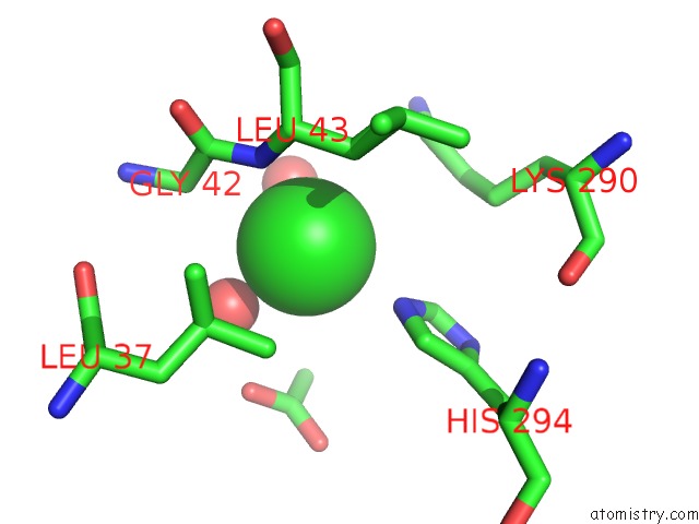
Mono view
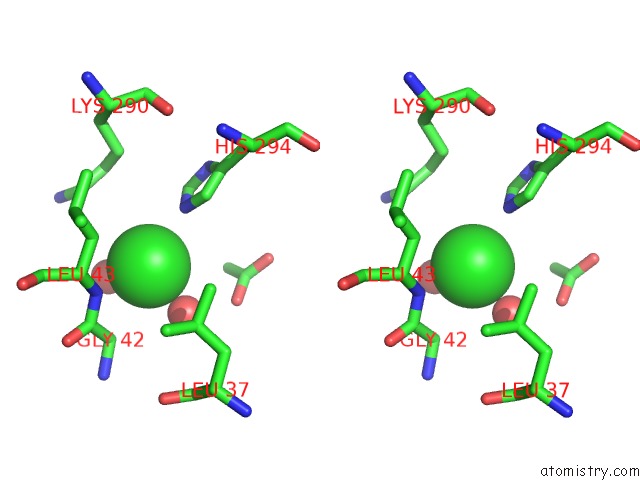
Stereo pair view
Mono view
Stereo pair view
A full contact list of Chlorine with other atoms in the Cl binding
site number 1 of Human [Protein Adp-Ribosylargenine] Hydrolase ARH1 in Complex with Adp-Hpm within 5.0Å range:
|
Reference:
J.G.M.Rack,
A.Ariza,
B.S.Drown,
C.Henfrey,
E.Bartlett,
T.Shirai,
P.J.Hergenrother,
I.Ahel.
(Adp-Ribosyl)Hydrolases: Structural Basis For Differential Substrate Recognition and Inhibition. Cell Chem Biol V. 25 1533 2018.
ISSN: ESSN 2451-9448
PubMed: 30472116
DOI: 10.1016/J.CHEMBIOL.2018.11.001
Page generated: Sat Jul 27 23:40:37 2024
ISSN: ESSN 2451-9448
PubMed: 30472116
DOI: 10.1016/J.CHEMBIOL.2018.11.001
Last articles
Zn in 9J0NZn in 9J0O
Zn in 9J0P
Zn in 9FJX
Zn in 9EKB
Zn in 9C0F
Zn in 9CAH
Zn in 9CH0
Zn in 9CH3
Zn in 9CH1