Chlorine »
PDB 6g1v-6g8l »
6g7r »
Chlorine in PDB 6g7r: Structure of Fully Reduced Variant E28Q of E. Coli Hydrogenase-1 at pH 8
Enzymatic activity of Structure of Fully Reduced Variant E28Q of E. Coli Hydrogenase-1 at pH 8
All present enzymatic activity of Structure of Fully Reduced Variant E28Q of E. Coli Hydrogenase-1 at pH 8:
1.12.99.6;
1.12.99.6;
Protein crystallography data
The structure of Structure of Fully Reduced Variant E28Q of E. Coli Hydrogenase-1 at pH 8, PDB code: 6g7r
was solved by
S.B.Carr,
F.A.Armstrong,
R.M.Evans,
with X-Ray Crystallography technique. A brief refinement statistics is given in the table below:
| Resolution Low / High (Å) | 67.89 / 1.20 |
| Space group | P 21 21 21 |
| Cell size a, b, c (Å), α, β, γ (°) | 94.068, 97.814, 183.210, 90.00, 90.00, 90.00 |
| R / Rfree (%) | 11.1 / 13.3 |
Other elements in 6g7r:
The structure of Structure of Fully Reduced Variant E28Q of E. Coli Hydrogenase-1 at pH 8 also contains other interesting chemical elements:
| Nickel | (Ni) | 2 atoms |
| Magnesium | (Mg) | 2 atoms |
| Iron | (Fe) | 24 atoms |
Chlorine Binding Sites:
The binding sites of Chlorine atom in the Structure of Fully Reduced Variant E28Q of E. Coli Hydrogenase-1 at pH 8
(pdb code 6g7r). This binding sites where shown within
5.0 Angstroms radius around Chlorine atom.
In total 4 binding sites of Chlorine where determined in the Structure of Fully Reduced Variant E28Q of E. Coli Hydrogenase-1 at pH 8, PDB code: 6g7r:
Jump to Chlorine binding site number: 1; 2; 3; 4;
In total 4 binding sites of Chlorine where determined in the Structure of Fully Reduced Variant E28Q of E. Coli Hydrogenase-1 at pH 8, PDB code: 6g7r:
Jump to Chlorine binding site number: 1; 2; 3; 4;
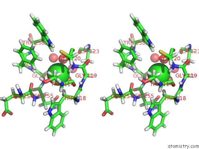
Chlorine binding site 1 out of 4 in 6g7r
Go back to
Chlorine binding site 1 out
of 4 in the Structure of Fully Reduced Variant E28Q of E. Coli Hydrogenase-1 at pH 8
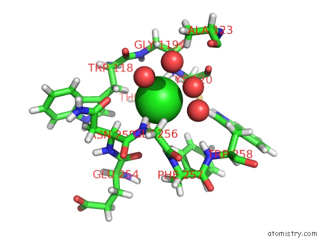
Mono view
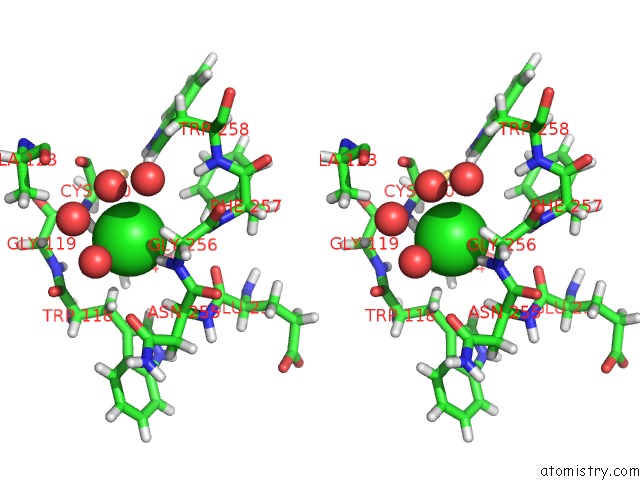
Stereo pair view
Mono view
Stereo pair view
A full contact list of Chlorine with other atoms in the Cl binding
site number 1 of Structure of Fully Reduced Variant E28Q of E. Coli Hydrogenase-1 at pH 8 within 5.0Å range:
|
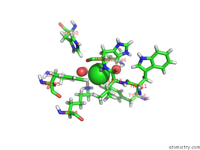
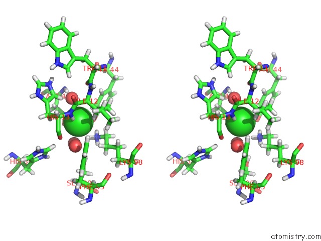
Chlorine binding site 2 out of 4 in 6g7r
Go back to
Chlorine binding site 2 out
of 4 in the Structure of Fully Reduced Variant E28Q of E. Coli Hydrogenase-1 at pH 8
Mono view
Stereo pair view
Mono view
Stereo pair view
A full contact list of Chlorine with other atoms in the Cl binding
site number 2 of Structure of Fully Reduced Variant E28Q of E. Coli Hydrogenase-1 at pH 8 within 5.0Å range:
|
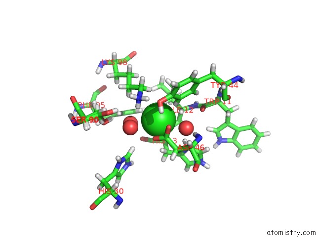
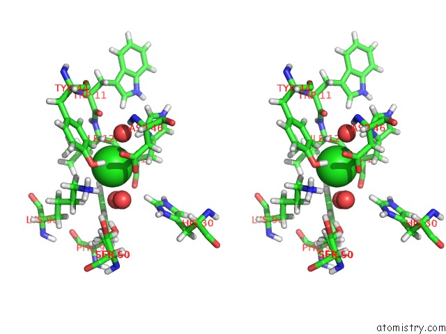
Chlorine binding site 3 out of 4 in 6g7r
Go back to
Chlorine binding site 3 out
of 4 in the Structure of Fully Reduced Variant E28Q of E. Coli Hydrogenase-1 at pH 8

Mono view
Stereo pair view
Mono view
Stereo pair view
A full contact list of Chlorine with other atoms in the Cl binding
site number 3 of Structure of Fully Reduced Variant E28Q of E. Coli Hydrogenase-1 at pH 8 within 5.0Å range:
|
Chlorine binding site 4 out of 4 in 6g7r
Go back to
Chlorine binding site 4 out
of 4 in the Structure of Fully Reduced Variant E28Q of E. Coli Hydrogenase-1 at pH 8
Mono view
Stereo pair view
Mono view
Stereo pair view
A full contact list of Chlorine with other atoms in the Cl binding
site number 4 of Structure of Fully Reduced Variant E28Q of E. Coli Hydrogenase-1 at pH 8 within 5.0Å range:
|
Reference:
R.M.Evans,
P.A.Ash,
S.E.Beaton,
E.J.Brooke,
K.A.Vincent,
S.B.Carr,
F.A.Armstrong.
Mechanistic Exploitation of A Self-Repairing, Blocked Proton Transfer Pathway in An O2-Tolerant [Nife]-Hydrogenase. J. Am. Chem. Soc. V. 140 10208 2018.
ISSN: ESSN 1520-5126
PubMed: 30070475
DOI: 10.1021/JACS.8B04798
Page generated: Sat Jul 27 23:49:06 2024
ISSN: ESSN 1520-5126
PubMed: 30070475
DOI: 10.1021/JACS.8B04798
Last articles
Ca in 5MSTCa in 5MS4
Ca in 5MS9
Ca in 5MQR
Ca in 5MQS
Ca in 5MS3
Ca in 5MQM
Ca in 5MQN
Ca in 5MPY
Ca in 5MOR