Chlorine »
PDB 6g8m-6ggn »
6g8p »
Chlorine in PDB 6g8p: 14-3-3SIGMA in Complex with A P129BETA3P and L132BETA3L Mutated Yap PS127 Phosphopeptide
Protein crystallography data
The structure of 14-3-3SIGMA in Complex with A P129BETA3P and L132BETA3L Mutated Yap PS127 Phosphopeptide, PDB code: 6g8p
was solved by
S.A.Andrei,
V.Thijssen,
L.Brunsveld,
C.Ottmann,
L.G.Milroy,
with X-Ray Crystallography technique. A brief refinement statistics is given in the table below:
| Resolution Low / High (Å) | 41.59 / 1.90 |
| Space group | C 2 2 21 |
| Cell size a, b, c (Å), α, β, γ (°) | 82.037, 111.499, 62.460, 90.00, 90.00, 90.00 |
| R / Rfree (%) | 13.3 / 15.9 |
Other elements in 6g8p:
The structure of 14-3-3SIGMA in Complex with A P129BETA3P and L132BETA3L Mutated Yap PS127 Phosphopeptide also contains other interesting chemical elements:
| Calcium | (Ca) | 2 atoms |
Chlorine Binding Sites:
The binding sites of Chlorine atom in the 14-3-3SIGMA in Complex with A P129BETA3P and L132BETA3L Mutated Yap PS127 Phosphopeptide
(pdb code 6g8p). This binding sites where shown within
5.0 Angstroms radius around Chlorine atom.
In total only one binding site of Chlorine was determined in the 14-3-3SIGMA in Complex with A P129BETA3P and L132BETA3L Mutated Yap PS127 Phosphopeptide, PDB code: 6g8p:
In total only one binding site of Chlorine was determined in the 14-3-3SIGMA in Complex with A P129BETA3P and L132BETA3L Mutated Yap PS127 Phosphopeptide, PDB code: 6g8p:
Chlorine binding site 1 out of 1 in 6g8p
Go back to
Chlorine binding site 1 out
of 1 in the 14-3-3SIGMA in Complex with A P129BETA3P and L132BETA3L Mutated Yap PS127 Phosphopeptide
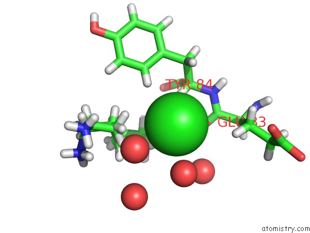
Mono view
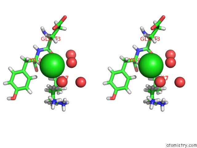
Stereo pair view
Mono view
Stereo pair view
A full contact list of Chlorine with other atoms in the Cl binding
site number 1 of 14-3-3SIGMA in Complex with A P129BETA3P and L132BETA3L Mutated Yap PS127 Phosphopeptide within 5.0Å range:
|
Reference:
S.A.Andrei,
V.Thijssen,
L.Brunsveld,
C.Ottmann,
L.G.Milroy.
A Study on the Effect of Synthetic Alpha-to-BETA3-Amino Acid Mutations on the Binding of Phosphopeptides to 14-3-3 Proteins. Chem.Commun.(Camb.) 2019.
ISSN: ESSN 1364-548X
PubMed: 31763628
DOI: 10.1039/C9CC07982C
Page generated: Sat Jul 12 14:27:15 2025
ISSN: ESSN 1364-548X
PubMed: 31763628
DOI: 10.1039/C9CC07982C
Last articles
Fe in 2YXOFe in 2YRS
Fe in 2YXC
Fe in 2YNM
Fe in 2YVJ
Fe in 2YP1
Fe in 2YU2
Fe in 2YU1
Fe in 2YQB
Fe in 2YOO