Chlorine »
PDB 6gp4-6gxd »
6gus »
Chlorine in PDB 6gus: Crystal Structure of Protein E From Non-Typeable Haemophilus Influenzae
Protein crystallography data
The structure of Crystal Structure of Protein E From Non-Typeable Haemophilus Influenzae, PDB code: 6gus
was solved by
D.Somers,
with X-Ray Crystallography technique. A brief refinement statistics is given in the table below:
| Resolution Low / High (Å) | 20.00 / 1.92 |
| Space group | I 41 |
| Cell size a, b, c (Å), α, β, γ (°) | 77.670, 77.670, 66.130, 90.00, 90.00, 90.00 |
| R / Rfree (%) | 18.5 / 24.4 |
Chlorine Binding Sites:
The binding sites of Chlorine atom in the Crystal Structure of Protein E From Non-Typeable Haemophilus Influenzae
(pdb code 6gus). This binding sites where shown within
5.0 Angstroms radius around Chlorine atom.
In total only one binding site of Chlorine was determined in the Crystal Structure of Protein E From Non-Typeable Haemophilus Influenzae, PDB code: 6gus:
In total only one binding site of Chlorine was determined in the Crystal Structure of Protein E From Non-Typeable Haemophilus Influenzae, PDB code: 6gus:
Chlorine binding site 1 out of 1 in 6gus
Go back to
Chlorine binding site 1 out
of 1 in the Crystal Structure of Protein E From Non-Typeable Haemophilus Influenzae
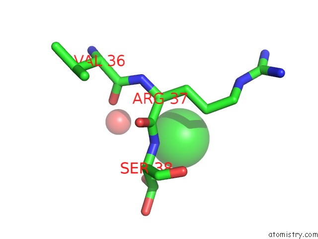
Mono view
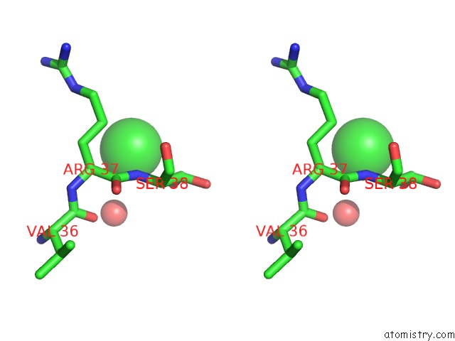
Stereo pair view
Mono view
Stereo pair view
A full contact list of Chlorine with other atoms in the Cl binding
site number 1 of Crystal Structure of Protein E From Non-Typeable Haemophilus Influenzae within 5.0Å range:
|
Reference:
N.Blais,
D.Somers,
D.Faubert,
S.Labbe,
C.Castado,
C.Ysebaert,
L.P.Gagnon,
J.Champagne,
M.Gagne,
D.Martin.
Design and Characterization of Protein E-Pila, A Candidate Fusion Antigen For Nontypeable Haemophilus Influenzae Vaccine. Infect.Immun. V. 87 2019.
ISSN: ESSN 1098-5522
PubMed: 31085711
DOI: 10.1128/IAI.00022-19
Page generated: Sun Jul 28 00:14:00 2024
ISSN: ESSN 1098-5522
PubMed: 31085711
DOI: 10.1128/IAI.00022-19
Last articles
Zn in 9J0NZn in 9J0O
Zn in 9J0P
Zn in 9FJX
Zn in 9EKB
Zn in 9C0F
Zn in 9CAH
Zn in 9CH0
Zn in 9CH3
Zn in 9CH1