Chlorine »
PDB 6gxj-6h4f »
6gxj »
Chlorine in PDB 6gxj: X-Ray Structure of Diru-1-Encapsulated Apoferritin
Protein crystallography data
The structure of X-Ray Structure of Diru-1-Encapsulated Apoferritin, PDB code: 6gxj
was solved by
A.Pica,
G.Ferraro,
A.Merlino,
with X-Ray Crystallography technique. A brief refinement statistics is given in the table below:
| Resolution Low / High (Å) | 14.26 / 1.43 |
| Space group | F 4 3 2 |
| Cell size a, b, c (Å), α, β, γ (°) | 180.404, 180.404, 180.404, 90.00, 90.00, 90.00 |
| R / Rfree (%) | 17.4 / 19.1 |
Other elements in 6gxj:
The structure of X-Ray Structure of Diru-1-Encapsulated Apoferritin also contains other interesting chemical elements:
| Cadmium | (Cd) | 10 atoms |
Chlorine Binding Sites:
The binding sites of Chlorine atom in the X-Ray Structure of Diru-1-Encapsulated Apoferritin
(pdb code 6gxj). This binding sites where shown within
5.0 Angstroms radius around Chlorine atom.
In total 2 binding sites of Chlorine where determined in the X-Ray Structure of Diru-1-Encapsulated Apoferritin, PDB code: 6gxj:
Jump to Chlorine binding site number: 1; 2;
In total 2 binding sites of Chlorine where determined in the X-Ray Structure of Diru-1-Encapsulated Apoferritin, PDB code: 6gxj:
Jump to Chlorine binding site number: 1; 2;
Chlorine binding site 1 out of 2 in 6gxj
Go back to
Chlorine binding site 1 out
of 2 in the X-Ray Structure of Diru-1-Encapsulated Apoferritin
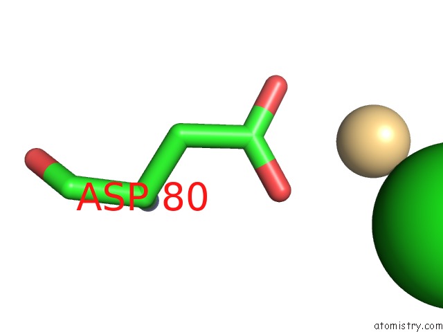
Mono view
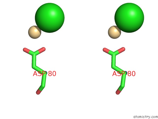
Stereo pair view
Mono view
Stereo pair view
A full contact list of Chlorine with other atoms in the Cl binding
site number 1 of X-Ray Structure of Diru-1-Encapsulated Apoferritin within 5.0Å range:
|
Chlorine binding site 2 out of 2 in 6gxj
Go back to
Chlorine binding site 2 out
of 2 in the X-Ray Structure of Diru-1-Encapsulated Apoferritin
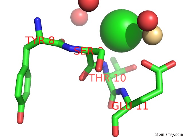
Mono view
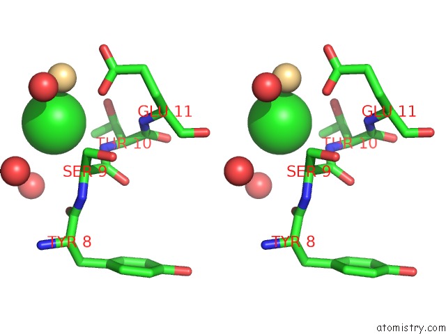
Stereo pair view
Mono view
Stereo pair view
A full contact list of Chlorine with other atoms in the Cl binding
site number 2 of X-Ray Structure of Diru-1-Encapsulated Apoferritin within 5.0Å range:
|
Reference:
G.Petruk,
D.M.Monti,
G.Ferraro,
A.Pica,
L.D'elia,
F.Pane,
A.Amoresano,
J.Furrer,
K.Kowalski,
A.Merlino.
Encapsulation of the Dinuclear Trithiolato-Bridged Arene Ruthenium Complex Diruthenium-1 in An Apoferritin Nanocage: Structure and Cytotoxicity. Chemmedchem V. 14 594 2019.
ISSN: ESSN 1860-7187
PubMed: 30674089
DOI: 10.1002/CMDC.201800805
Page generated: Sun Jul 28 00:20:41 2024
ISSN: ESSN 1860-7187
PubMed: 30674089
DOI: 10.1002/CMDC.201800805
Last articles
Zn in 9J0NZn in 9J0O
Zn in 9J0P
Zn in 9FJX
Zn in 9EKB
Zn in 9C0F
Zn in 9CAH
Zn in 9CH0
Zn in 9CH3
Zn in 9CH1