Chlorine »
PDB 6gxj-6h4f »
6h3x »
Chlorine in PDB 6h3x: Oropouche Virus Glycoprotein Gc Head Domain
Protein crystallography data
The structure of Oropouche Virus Glycoprotein Gc Head Domain, PDB code: 6h3x
was solved by
J.Hellert,
A.Aebischer,
K.Wernike,
A.Haouz,
E.Brocchi,
S.Reiche,
P.Guardado-Calvo,
M.Beer,
F.A.Rey,
with X-Ray Crystallography technique. A brief refinement statistics is given in the table below:
| Resolution Low / High (Å) | 33.90 / 2.09 |
| Space group | H 3 2 |
| Cell size a, b, c (Å), α, β, γ (°) | 104.638, 104.638, 133.504, 90.00, 90.00, 120.00 |
| R / Rfree (%) | 20.3 / 23.3 |
Chlorine Binding Sites:
The binding sites of Chlorine atom in the Oropouche Virus Glycoprotein Gc Head Domain
(pdb code 6h3x). This binding sites where shown within
5.0 Angstroms radius around Chlorine atom.
In total 2 binding sites of Chlorine where determined in the Oropouche Virus Glycoprotein Gc Head Domain, PDB code: 6h3x:
Jump to Chlorine binding site number: 1; 2;
In total 2 binding sites of Chlorine where determined in the Oropouche Virus Glycoprotein Gc Head Domain, PDB code: 6h3x:
Jump to Chlorine binding site number: 1; 2;
Chlorine binding site 1 out of 2 in 6h3x
Go back to
Chlorine binding site 1 out
of 2 in the Oropouche Virus Glycoprotein Gc Head Domain
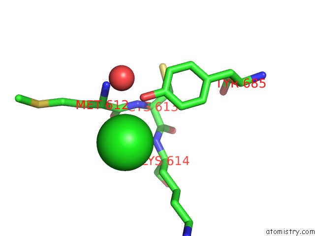
Mono view
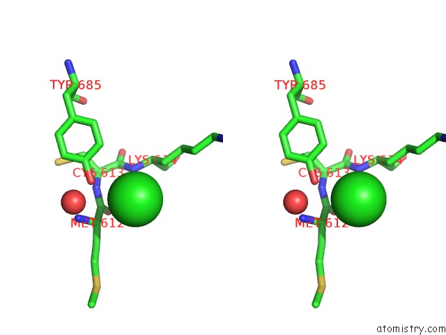
Stereo pair view
Mono view
Stereo pair view
A full contact list of Chlorine with other atoms in the Cl binding
site number 1 of Oropouche Virus Glycoprotein Gc Head Domain within 5.0Å range:
|
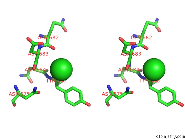
Chlorine binding site 2 out of 2 in 6h3x
Go back to
Chlorine binding site 2 out
of 2 in the Oropouche Virus Glycoprotein Gc Head Domain
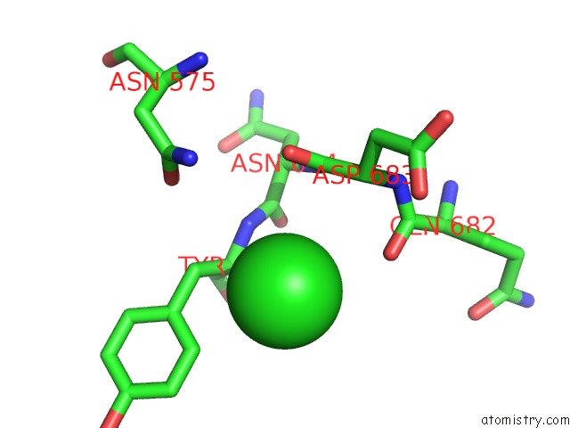
Mono view
Stereo pair view
Mono view
Stereo pair view
A full contact list of Chlorine with other atoms in the Cl binding
site number 2 of Oropouche Virus Glycoprotein Gc Head Domain within 5.0Å range:
|
Reference:
J.Hellert,
A.Aebischer,
K.Wernike,
A.Haouz,
E.Brocchi,
S.Reiche,
P.Guardado-Calvo,
M.Beer,
F.A.Rey.
Orthobunyavirus Spike Architecture and Recognition By Neutralizing Antibodies. Nat Commun V. 10 879 2019.
ISSN: ESSN 2041-1723
PubMed: 30787296
DOI: 10.1038/S41467-019-08832-8
Page generated: Sun Jul 28 00:27:22 2024
ISSN: ESSN 2041-1723
PubMed: 30787296
DOI: 10.1038/S41467-019-08832-8
Last articles
Zn in 9J0NZn in 9J0O
Zn in 9J0P
Zn in 9FJX
Zn in 9EKB
Zn in 9C0F
Zn in 9CAH
Zn in 9CH0
Zn in 9CH3
Zn in 9CH1