Chlorine »
PDB 6hb5-6hkj »
6hcc »
Chlorine in PDB 6hcc: Structure of GLUA2 Ligand-Binding Domain (S1S2J-N775S) in Complex with Glutamate and TDPAM02 at 1.6 A Resolution.
Protein crystallography data
The structure of Structure of GLUA2 Ligand-Binding Domain (S1S2J-N775S) in Complex with Glutamate and TDPAM02 at 1.6 A Resolution., PDB code: 6hcc
was solved by
S.Laulumaa,
K.V.Hansen,
K.Frydenvang,
J.S.Kastrup,
with X-Ray Crystallography technique. A brief refinement statistics is given in the table below:
| Resolution Low / High (Å) | 29.74 / 1.62 |
| Space group | P 21 21 2 |
| Cell size a, b, c (Å), α, β, γ (°) | 97.979, 121.967, 47.395, 90.00, 90.00, 90.00 |
| R / Rfree (%) | 15.5 / 18.2 |
Chlorine Binding Sites:
The binding sites of Chlorine atom in the Structure of GLUA2 Ligand-Binding Domain (S1S2J-N775S) in Complex with Glutamate and TDPAM02 at 1.6 A Resolution.
(pdb code 6hcc). This binding sites where shown within
5.0 Angstroms radius around Chlorine atom.
In total 3 binding sites of Chlorine where determined in the Structure of GLUA2 Ligand-Binding Domain (S1S2J-N775S) in Complex with Glutamate and TDPAM02 at 1.6 A Resolution., PDB code: 6hcc:
Jump to Chlorine binding site number: 1; 2; 3;
In total 3 binding sites of Chlorine where determined in the Structure of GLUA2 Ligand-Binding Domain (S1S2J-N775S) in Complex with Glutamate and TDPAM02 at 1.6 A Resolution., PDB code: 6hcc:
Jump to Chlorine binding site number: 1; 2; 3;
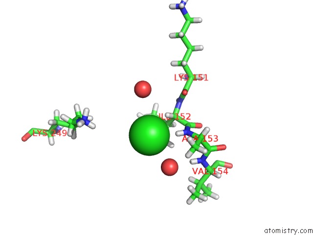
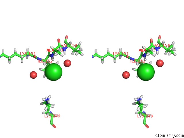
Chlorine binding site 1 out of 3 in 6hcc
Go back to
Chlorine binding site 1 out
of 3 in the Structure of GLUA2 Ligand-Binding Domain (S1S2J-N775S) in Complex with Glutamate and TDPAM02 at 1.6 A Resolution.
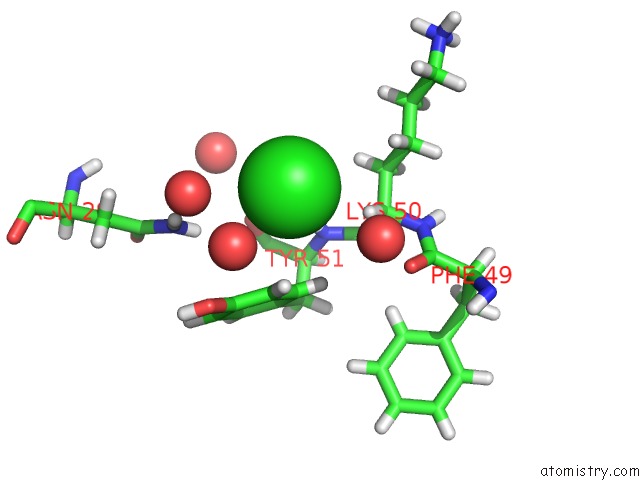
Mono view
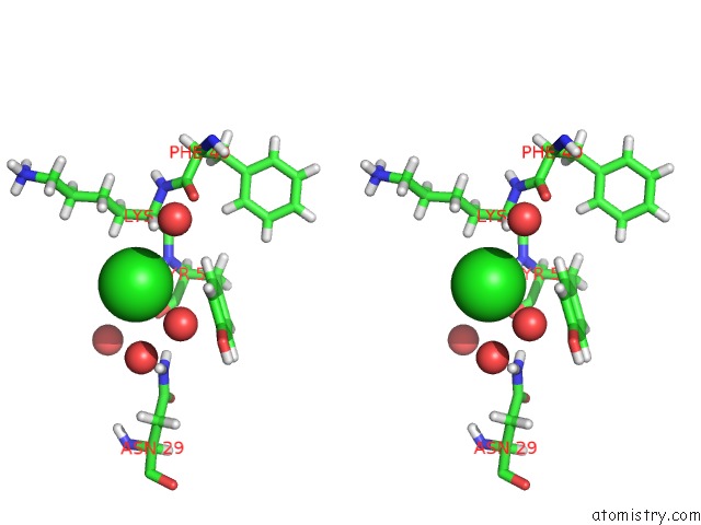
Stereo pair view
Mono view
Stereo pair view
A full contact list of Chlorine with other atoms in the Cl binding
site number 1 of Structure of GLUA2 Ligand-Binding Domain (S1S2J-N775S) in Complex with Glutamate and TDPAM02 at 1.6 A Resolution. within 5.0Å range:
|
Chlorine binding site 2 out of 3 in 6hcc
Go back to
Chlorine binding site 2 out
of 3 in the Structure of GLUA2 Ligand-Binding Domain (S1S2J-N775S) in Complex with Glutamate and TDPAM02 at 1.6 A Resolution.
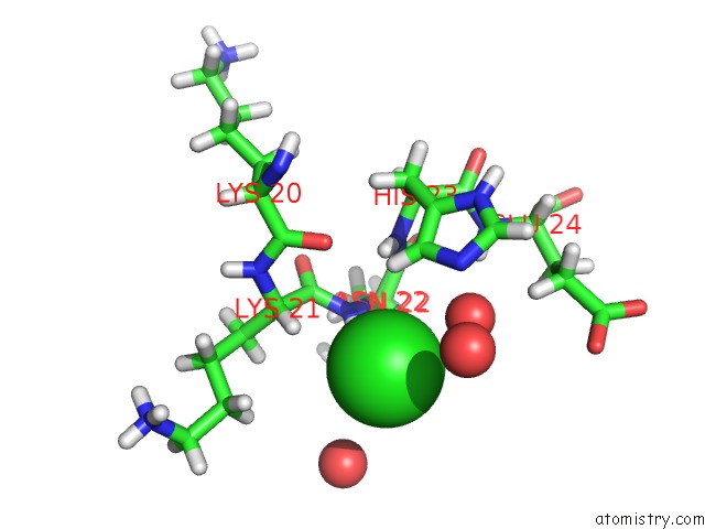
Mono view
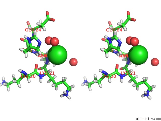
Stereo pair view
Mono view
Stereo pair view
A full contact list of Chlorine with other atoms in the Cl binding
site number 2 of Structure of GLUA2 Ligand-Binding Domain (S1S2J-N775S) in Complex with Glutamate and TDPAM02 at 1.6 A Resolution. within 5.0Å range:
|
Chlorine binding site 3 out of 3 in 6hcc
Go back to
Chlorine binding site 3 out
of 3 in the Structure of GLUA2 Ligand-Binding Domain (S1S2J-N775S) in Complex with Glutamate and TDPAM02 at 1.6 A Resolution.
Mono view
Stereo pair view
Mono view
Stereo pair view
A full contact list of Chlorine with other atoms in the Cl binding
site number 3 of Structure of GLUA2 Ligand-Binding Domain (S1S2J-N775S) in Complex with Glutamate and TDPAM02 at 1.6 A Resolution. within 5.0Å range:
|
Reference:
S.Laulumaa,
K.V.Hansen,
M.Masternak,
T.Drapier,
P.Francotte,
B.Pirotte,
K.Frydenvang,
J.S.Kastrup.
Crystal Structures of Potent Dimeric Positive Allosteric Modulators at the Ligand-Binding Domain of the GLUA2 Receptor. Acs Med.Chem.Lett. V. 10 243 2019.
ISSN: ISSN 1948-5875
PubMed: 30891120
DOI: 10.1021/ACSMEDCHEMLETT.8B00369
Page generated: Sun Jul 28 00:45:47 2024
ISSN: ISSN 1948-5875
PubMed: 30891120
DOI: 10.1021/ACSMEDCHEMLETT.8B00369
Last articles
Zn in 9J0NZn in 9J0O
Zn in 9J0P
Zn in 9FJX
Zn in 9EKB
Zn in 9C0F
Zn in 9CAH
Zn in 9CH0
Zn in 9CH3
Zn in 9CH1