Chlorine in PDB 6hrr: Structure of the TRPML2 Eld at pH 6.5

Protein crystallography data

The structure of Structure of the TRPML2 Eld at pH 6.5, PDB code: 6hrr was solved by N.Bader, K.K.Viet, A.Wagner, U.A.Hellmich, with X-Ray Crystallography technique. A brief refinement statistics is given in the table below:

Resolution Low / High (Å) 20.00 / 2.00
Space group I 4 2 2
Cell size a, b, c (Å), α, β, γ (°) 109.471, 109.471, 149.744, 90.00, 90.00, 90.00
R / Rfree (%) 18.6 / 21.7

Other elements in 6hrr:

The structure of Structure of the TRPML2 Eld at pH 6.5 also contains other interesting chemical elements:

Potassium (K) 1 atom

Chlorine Binding Sites:

The binding sites of Chlorine atom in the Structure of the TRPML2 Eld at pH 6.5 (pdb code 6hrr). This binding sites where shown within 5.0 Angstroms radius around Chlorine atom.
In total 3 binding sites of Chlorine where determined in the Structure of the TRPML2 Eld at pH 6.5, PDB code: 6hrr:
Jump to Chlorine binding site number: 1; 2; 3;

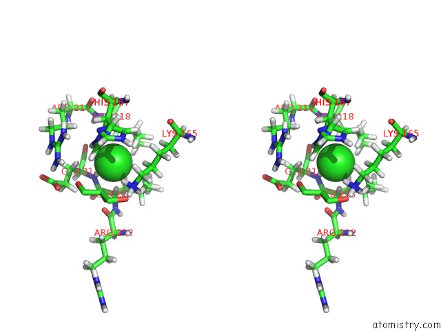
Chlorine binding site 1 out of 3 in 6hrr

Go back to Chlorine Binding Sites List in 6hrr
Chlorine binding site 1 out of 3 in the Structure of the TRPML2 Eld at pH 6.5


Mono view


Stereo pair view

A full contact list of Chlorine with other atoms in the Cl binding site number 1 of Structure of the TRPML2 Eld at pH 6.5 within 5.0Å range:
probe atom residue distance (Å) B Occ
A:Cl302

b:57.6
occ:1.00
H A:GLU214 2.4 54.2 0.0
HZ2 A:LYS165 2.5 55.7 0.0
HA A:LEU213 2.9 49.5 0.0
HD13 A:LEU218 2.9 50.3 0.0
HD1 A:HIS167 2.9 72.8 0.0
HD2 A:LYS165 3.0 49.2 0.0
N A:GLU214 3.4 53.9 1.0
NZ A:LYS165 3.4 54.6 1.0
ND1 A:HIS167 3.4 72.6 0.4
HD22 A:LEU213 3.5 47.9 0.0
HB2 A:GLU214 3.5 58.2 0.0
HB3 A:HIS167 3.5 62.1 0.0
HB3 A:HIS167 3.6 63.4 0.0
HB2 A:LEU218 3.6 51.4 0.0
HE3 A:LYS165 3.7 54.9 0.0
HZ3 A:LYS165 3.7 54.1 0.0
HD23 A:LEU213 3.8 48.7 0.0
CA A:LEU213 3.8 49.0 1.0
CE A:LYS165 3.9 53.4 1.0
CD A:LYS165 3.9 49.5 1.0
CD1 A:LEU218 3.9 50.2 1.0
HB3 A:LEU213 4.0 47.1 0.0
HD12 A:LEU218 4.0 51.1 0.0
CG A:HIS167 4.1 69.8 0.4
C A:LEU213 4.1 55.7 1.0
CD2 A:LEU213 4.1 48.0 1.0
HZ1 A:LYS165 4.1 56.2 0.0
CE1 A:HIS167 4.2 73.2 0.4
CB A:HIS167 4.2 64.4 0.4
O A:GLU214 4.2 55.1 1.0
CB A:HIS167 4.3 63.1 0.6
HB2 A:HIS167 4.3 65.8 0.0
CG A:HIS167 4.3 67.3 0.6
CA A:GLU214 4.3 54.3 1.0
CB A:GLU214 4.3 58.1 1.0
HB2 A:HIS167 4.4 64.7 0.0
HG3 A:GLU214 4.4 73.8 0.0
O2 A:GOL307 4.4 93.1 0.5
O A:ARG212 4.4 58.0 1.0
CB A:LEU213 4.4 46.9 1.0
HD22 A:LEU218 4.4 55.9 0.0
HE1 A:HIS167 4.4 74.1 0.0
HA A:LEU218 4.4 52.8 0.0
CD2 A:HIS167 4.4 68.2 0.6
CB A:LEU218 4.5 51.1 1.0
HD3 A:LYS165 4.5 48.0 0.0
HD11 A:LEU218 4.6 49.0 0.0
HD2 A:HIS167 4.6 67.5 0.0
C A:GLU214 4.6 55.6 1.0
HG3 A:LYS165 4.6 52.4 0.0
CG A:LEU218 4.7 51.9 1.0
O3 A:GOL307 4.8 94.9 0.5
HB2 A:LYS165 4.8 48.3 0.0
HG3 A:ARG217 4.8 65.9 0.0
CG A:LYS165 4.9 50.8 1.0
ND1 A:HIS167 4.9 71.2 0.6
CG A:GLU214 4.9 74.7 1.0
N A:LEU213 4.9 49.3 1.0
CG A:LEU213 4.9 48.9 1.0
C2 A:GOL307 4.9 93.6 0.5
HE2 A:LYS165 5.0 53.6 0.0
HD21 A:LEU213 5.0 46.9 0.0
CD2 A:HIS167 5.0 73.1 0.4
CA A:LEU218 5.0 52.5 1.0

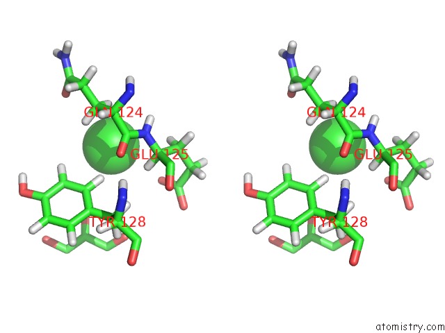
Chlorine binding site 2 out of 3 in 6hrr

Go back to Chlorine Binding Sites List in 6hrr
Chlorine binding site 2 out of 3 in the Structure of the TRPML2 Eld at pH 6.5


Mono view


Stereo pair view

A full contact list of Chlorine with other atoms in the Cl binding site number 2 of Structure of the TRPML2 Eld at pH 6.5 within 5.0Å range:
probe atom residue distance (Å) B Occ
A:Cl303

b:1.0
occ:1.00
HB2 A:GLN124 3.5 43.2 0.0
HD2 A:TYR128 3.6 42.1 0.0
HA A:GLU125 3.6 43.2 0.0
OE1 A:GLU125 3.8 81.7 1.0
HB3 A:GLN124 3.8 42.6 0.0
HB2 A:GLU125 3.8 46.2 0.0
HE2 A:TYR128 3.9 44.0 0.0
N A:GLU125 4.0 43.2 1.0
H A:GLU125 4.1 44.2 0.0
CB A:GLN124 4.1 42.4 1.0
CA A:GLU125 4.2 43.0 1.0
C A:GLN124 4.4 43.9 1.0
OE1 A:GLN124 4.4 47.2 1.0
CD2 A:TYR128 4.4 42.1 1.0
O2 A:GOL305 4.5 98.2 1.0
CB A:GLU125 4.5 45.3 1.0
CE2 A:TYR128 4.6 43.2 1.0
O A:GLN124 4.8 41.3 1.0
CD A:GLU125 4.8 86.5 1.0
CA A:GLN124 4.9 41.0 1.0

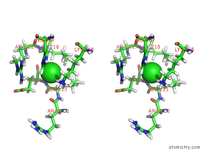
Chlorine binding site 3 out of 3 in 6hrr

Go back to Chlorine Binding Sites List in 6hrr
Chlorine binding site 3 out of 3 in the Structure of the TRPML2 Eld at pH 6.5


Mono view


Stereo pair view

A full contact list of Chlorine with other atoms in the Cl binding site number 3 of Structure of the TRPML2 Eld at pH 6.5 within 5.0Å range:
probe atom residue distance (Å) B Occ
B:Cl301

b:60.8
occ:1.00
HZ2 B:LYS165 2.2 53.9 0.0
H B:GLU214 2.3 53.9 0.0
HA B:LEU213 2.9 52.8 0.0
HD2 B:LYS165 3.0 47.0 0.0
HD13 B:LEU218 3.1 55.7 0.0
NZ B:LYS165 3.2 54.5 1.0
ND1 B:HIS167 3.2 66.2 1.0
N B:GLU214 3.3 54.1 1.0
HB2 B:GLU214 3.3 57.9 0.0
HE3 B:LYS165 3.5 55.2 0.0
HZ3 B:LYS165 3.6 53.3 0.0
HB3 B:HIS167 3.6 57.0 0.0
HB2 B:LEU218 3.7 51.7 0.0
HD22 B:LEU213 3.7 50.4 0.0
CE B:LYS165 3.7 54.7 1.0
CD B:LYS165 3.8 47.5 1.0
HZ1 B:LYS165 3.9 55.2 0.0
HD23 B:LEU213 3.9 51.9 0.0
CA B:LEU213 3.9 52.5 1.0
HB2 B:HIS167 4.0 58.8 0.0
CG B:HIS167 4.0 63.2 1.0
CE1 B:HIS167 4.0 66.9 1.0
CD1 B:LEU218 4.1 54.9 1.0
C B:LEU213 4.1 58.5 1.0
HB3 B:LEU213 4.1 49.7 0.0
CB B:HIS167 4.1 57.8 1.0
O B:GLU214 4.1 57.6 1.0
CA B:GLU214 4.2 54.9 1.0
CB B:GLU214 4.2 58.1 1.0
HD12 B:LEU218 4.2 55.9 0.0
HE1 B:HIS167 4.2 67.8 0.0
CD2 B:LEU213 4.3 50.8 1.0
HG3 B:GLU214 4.3 70.8 0.0
O B:ARG212 4.4 65.7 1.0
C B:GLU214 4.4 57.7 1.0
HA B:LEU218 4.5 52.2 0.0
CB B:LEU213 4.5 49.9 1.0
HG3 B:ARG217 4.5 76.0 0.0
HD3 B:LYS165 4.5 46.9 0.0
HG3 B:LYS165 4.6 50.3 0.0
HD22 B:LEU218 4.6 56.9 0.0
CB B:LEU218 4.6 50.7 1.0
HD11 B:LEU218 4.7 54.4 0.0
HE2 B:LYS165 4.8 54.9 0.0
CG B:LYS165 4.8 49.8 1.0
CG B:GLU214 4.8 71.2 1.0
CG B:LEU218 4.9 54.4 1.0
HB2 B:LYS165 4.9 49.6 0.0
N B:LEU213 5.0 54.5 1.0

Reference:

K.K.Viet, A.Wagner, K.Schwickert, N.Hellwig, M.Brennich, N.Bader, T.Schirmeister, N.Morgner, H.Schindelin, U.A.Hellmich. Structure of the Human TRPML2 Ion Channel Extracytosolic/Lumenal Domain. Structure V. 27 1246 2019.
ISSN: ISSN 0969-2126
PubMed: 31178222
DOI: 10.1016/J.STR.2019.04.016
Page generated: Sat Dec 12 13:05:58 2020

Last articles

Zn in 8WB0
Zn in 8WAX
Zn in 8WAU
Zn in 8WAZ
Zn in 8WAY
Zn in 8WAV
Zn in 8WAW
Zn in 8WAT
Zn in 8W7M
Zn in 8WD3
© Copyright 2008-2020 by atomistry.com
Home   |    Site Map   |    Copyright   |    Contact us   |    Privacy