Chlorine »
PDB 6hzz-6i6c »
6hzz »
Chlorine in PDB 6hzz: Structure of Human D-Glucuronyl C5 Epimerase
Enzymatic activity of Structure of Human D-Glucuronyl C5 Epimerase
All present enzymatic activity of Structure of Human D-Glucuronyl C5 Epimerase:
5.1.3.17;
5.1.3.17;
Protein crystallography data
The structure of Structure of Human D-Glucuronyl C5 Epimerase, PDB code: 6hzz
was solved by
C.Debarnot,
Y.R.Monneau,
V.Roig-Zamboni,
C.Le Narvor,
A.Goulet,
F.Fadel,
R.R.Vives,
D.Bonnaffe,
H.Lortat-Jacob,
Y.Bourne,
with X-Ray Crystallography technique. A brief refinement statistics is given in the table below:
| Resolution Low / High (Å) | 43.83 / 2.52 |
| Space group | P 61 |
| Cell size a, b, c (Å), α, β, γ (°) | 99.820, 99.820, 262.970, 90.00, 90.00, 120.00 |
| R / Rfree (%) | 17.6 / 22.6 |
Other elements in 6hzz:
The structure of Structure of Human D-Glucuronyl C5 Epimerase also contains other interesting chemical elements:
| Calcium | (Ca) | 2 atoms |
Chlorine Binding Sites:
The binding sites of Chlorine atom in the Structure of Human D-Glucuronyl C5 Epimerase
(pdb code 6hzz). This binding sites where shown within
5.0 Angstroms radius around Chlorine atom.
In total 2 binding sites of Chlorine where determined in the Structure of Human D-Glucuronyl C5 Epimerase, PDB code: 6hzz:
Jump to Chlorine binding site number: 1; 2;
In total 2 binding sites of Chlorine where determined in the Structure of Human D-Glucuronyl C5 Epimerase, PDB code: 6hzz:
Jump to Chlorine binding site number: 1; 2;
Chlorine binding site 1 out of 2 in 6hzz
Go back to
Chlorine binding site 1 out
of 2 in the Structure of Human D-Glucuronyl C5 Epimerase
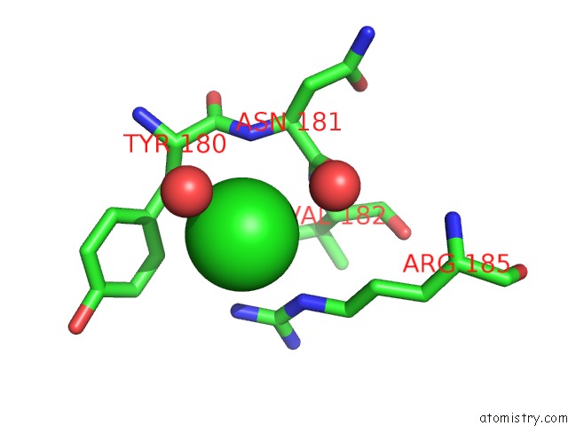
Mono view
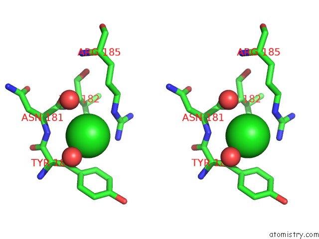
Stereo pair view
Mono view
Stereo pair view
A full contact list of Chlorine with other atoms in the Cl binding
site number 1 of Structure of Human D-Glucuronyl C5 Epimerase within 5.0Å range:
|
Chlorine binding site 2 out of 2 in 6hzz
Go back to
Chlorine binding site 2 out
of 2 in the Structure of Human D-Glucuronyl C5 Epimerase
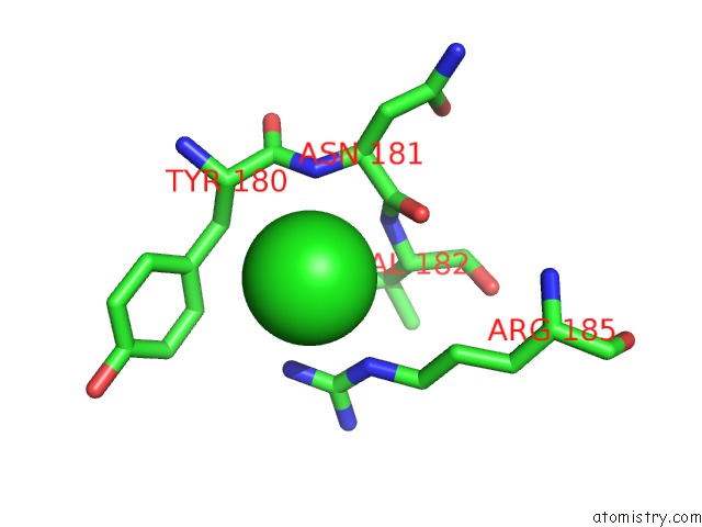
Mono view
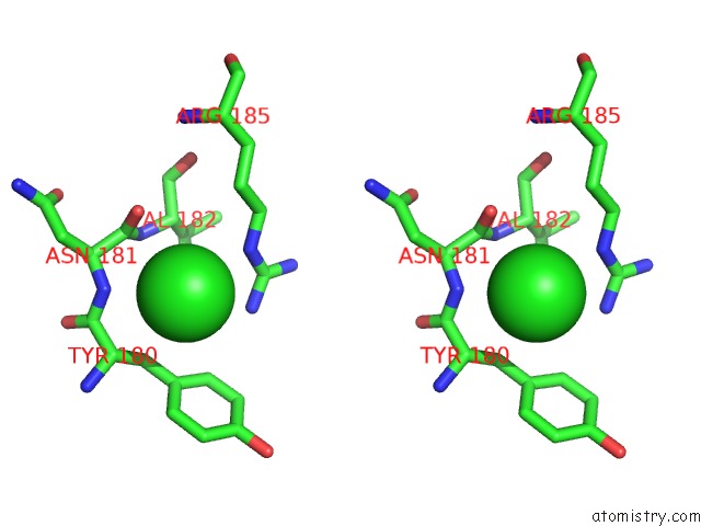
Stereo pair view
Mono view
Stereo pair view
A full contact list of Chlorine with other atoms in the Cl binding
site number 2 of Structure of Human D-Glucuronyl C5 Epimerase within 5.0Å range:
|
Reference:
C.Debarnot,
Y.R.Monneau,
V.Roig-Zamboni,
V.Delauzun,
C.Le Narvor,
E.Richard,
J.Henault,
A.Goulet,
F.Fadel,
R.R.Vives,
B.Priem,
D.Bonnaffe,
H.Lortat-Jacob,
Y.Bourne.
Substrate Binding Mode and Catalytic Mechanism of Human Heparan Sulfate D-Glucuronyl C5 Epimerase. Proc.Natl.Acad.Sci.Usa V. 116 6760 2019.
ISSN: ESSN 1091-6490
PubMed: 30872481
DOI: 10.1073/PNAS.1818333116
Page generated: Sun Jul 28 01:28:55 2024
ISSN: ESSN 1091-6490
PubMed: 30872481
DOI: 10.1073/PNAS.1818333116
Last articles
Zn in 9J0NZn in 9J0O
Zn in 9J0P
Zn in 9FJX
Zn in 9EKB
Zn in 9C0F
Zn in 9CAH
Zn in 9CH0
Zn in 9CH3
Zn in 9CH1