Chlorine »
PDB 6kkr-6kw1 »
6kvd »
Chlorine in PDB 6kvd: Crystal Structure of Human Nucleosome Containing H2A.J
Protein crystallography data
The structure of Crystal Structure of Human Nucleosome Containing H2A.J, PDB code: 6kvd
was solved by
H.Tanaka,
M.Koyama,
S.Sato,
T.Kujirai,
H.Kurumizaka,
with X-Ray Crystallography technique. A brief refinement statistics is given in the table below:
| Resolution Low / High (Å) | 47.84 / 2.21 |
| Space group | P 21 21 21 |
| Cell size a, b, c (Å), α, β, γ (°) | 99.640, 108.791, 171.177, 90.00, 90.00, 90.00 |
| R / Rfree (%) | 21.3 / 25.7 |
Other elements in 6kvd:
The structure of Crystal Structure of Human Nucleosome Containing H2A.J also contains other interesting chemical elements:
| Manganese | (Mn) | 13 atoms |
Chlorine Binding Sites:
The binding sites of Chlorine atom in the Crystal Structure of Human Nucleosome Containing H2A.J
(pdb code 6kvd). This binding sites where shown within
5.0 Angstroms radius around Chlorine atom.
In total 4 binding sites of Chlorine where determined in the Crystal Structure of Human Nucleosome Containing H2A.J, PDB code: 6kvd:
Jump to Chlorine binding site number: 1; 2; 3; 4;
In total 4 binding sites of Chlorine where determined in the Crystal Structure of Human Nucleosome Containing H2A.J, PDB code: 6kvd:
Jump to Chlorine binding site number: 1; 2; 3; 4;
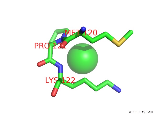
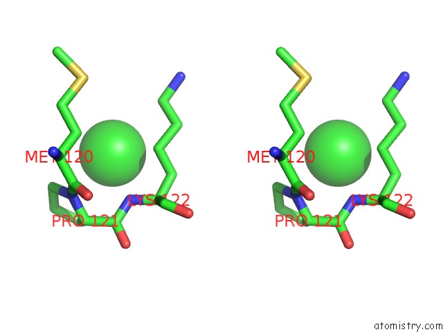
Chlorine binding site 1 out of 4 in 6kvd
Go back to
Chlorine binding site 1 out
of 4 in the Crystal Structure of Human Nucleosome Containing H2A.J
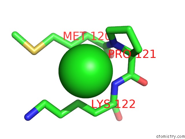
Mono view
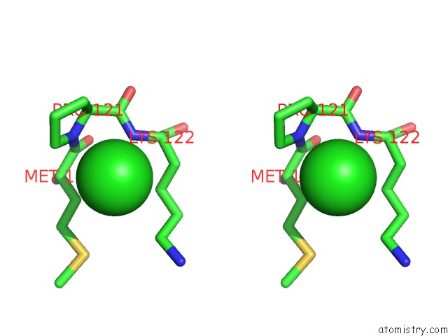
Stereo pair view
Mono view
Stereo pair view
A full contact list of Chlorine with other atoms in the Cl binding
site number 1 of Crystal Structure of Human Nucleosome Containing H2A.J within 5.0Å range:
|
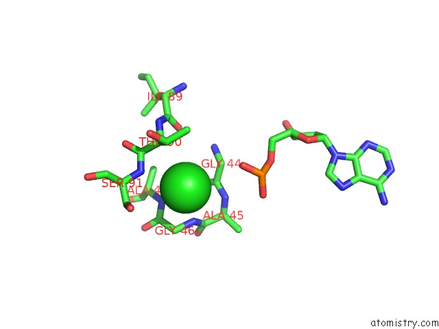
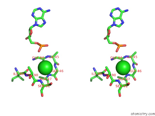
Chlorine binding site 2 out of 4 in 6kvd
Go back to
Chlorine binding site 2 out
of 4 in the Crystal Structure of Human Nucleosome Containing H2A.J
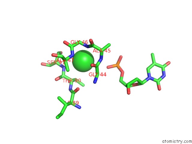
Mono view
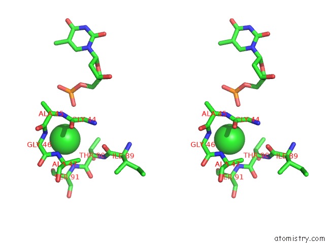
Stereo pair view
Mono view
Stereo pair view
A full contact list of Chlorine with other atoms in the Cl binding
site number 2 of Crystal Structure of Human Nucleosome Containing H2A.J within 5.0Å range:
|
Chlorine binding site 3 out of 4 in 6kvd
Go back to
Chlorine binding site 3 out
of 4 in the Crystal Structure of Human Nucleosome Containing H2A.J
Mono view
Stereo pair view
Mono view
Stereo pair view
A full contact list of Chlorine with other atoms in the Cl binding
site number 3 of Crystal Structure of Human Nucleosome Containing H2A.J within 5.0Å range:
|
Chlorine binding site 4 out of 4 in 6kvd
Go back to
Chlorine binding site 4 out
of 4 in the Crystal Structure of Human Nucleosome Containing H2A.J
Mono view
Stereo pair view
Mono view
Stereo pair view
A full contact list of Chlorine with other atoms in the Cl binding
site number 4 of Crystal Structure of Human Nucleosome Containing H2A.J within 5.0Å range:
|
Reference:
H.Tanaka,
S.Sato,
M.Koyama,
T.Kujirai,
H.Kurumizaka.
Biochemical and Structural Analyses of the Nucleosome Containing Human Histone H2A.J. J.Biochem. 2019.
ISSN: ISSN 0021-924X
PubMed: 31793981
DOI: 10.1093/JB/MVZ109
Page generated: Sun Jul 28 02:35:14 2024
ISSN: ISSN 0021-924X
PubMed: 31793981
DOI: 10.1093/JB/MVZ109
Last articles
Zn in 9J0NZn in 9J0O
Zn in 9J0P
Zn in 9FJX
Zn in 9EKB
Zn in 9C0F
Zn in 9CAH
Zn in 9CH0
Zn in 9CH3
Zn in 9CH1