Chlorine »
PDB 6lw2-6m7z »
6m0f »
Chlorine in PDB 6m0f: X-Ray Structure of Drosophila Dopamine Transporter with Subsiteb Mutations (D121G/S426M) in Substrate-Free Form
Protein crystallography data
The structure of X-Ray Structure of Drosophila Dopamine Transporter with Subsiteb Mutations (D121G/S426M) in Substrate-Free Form, PDB code: 6m0f
was solved by
P.Shabareesh,
A.K.Mallela,
D.Joseph,
A.Penmatsa,
with X-Ray Crystallography technique. A brief refinement statistics is given in the table below:
| Resolution Low / High (Å) | 48.99 / 3.30 |
| Space group | P 21 21 21 |
| Cell size a, b, c (Å), α, β, γ (°) | 97.978, 133.472, 162.088, 90, 90, 90 |
| R / Rfree (%) | 24.1 / 27.7 |
Other elements in 6m0f:
The structure of X-Ray Structure of Drosophila Dopamine Transporter with Subsiteb Mutations (D121G/S426M) in Substrate-Free Form also contains other interesting chemical elements:
| Sodium | (Na) | 2 atoms |
Chlorine Binding Sites:
The binding sites of Chlorine atom in the X-Ray Structure of Drosophila Dopamine Transporter with Subsiteb Mutations (D121G/S426M) in Substrate-Free Form
(pdb code 6m0f). This binding sites where shown within
5.0 Angstroms radius around Chlorine atom.
In total only one binding site of Chlorine was determined in the X-Ray Structure of Drosophila Dopamine Transporter with Subsiteb Mutations (D121G/S426M) in Substrate-Free Form, PDB code: 6m0f:
In total only one binding site of Chlorine was determined in the X-Ray Structure of Drosophila Dopamine Transporter with Subsiteb Mutations (D121G/S426M) in Substrate-Free Form, PDB code: 6m0f:
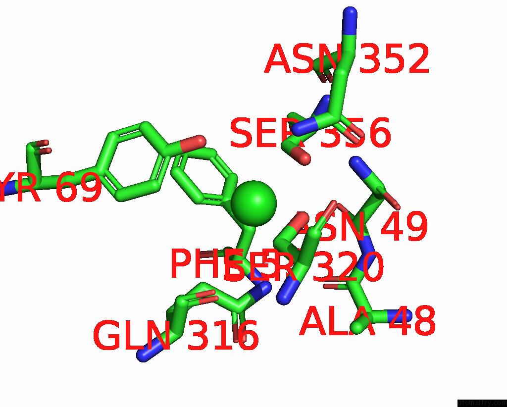
Chlorine binding site 1 out of 1 in 6m0f
Go back to
Chlorine binding site 1 out
of 1 in the X-Ray Structure of Drosophila Dopamine Transporter with Subsiteb Mutations (D121G/S426M) in Substrate-Free Form
Mono view
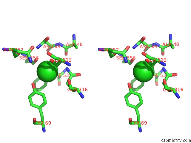
Stereo pair view
Mono view
Stereo pair view
A full contact list of Chlorine with other atoms in the Cl binding
site number 1 of X-Ray Structure of Drosophila Dopamine Transporter with Subsiteb Mutations (D121G/S426M) in Substrate-Free Form within 5.0Å range:
|
Reference:
P.Shabareesh,
A.K.Mallela,
D.Joseph.
Structural Basis of Norepinephrine Recognition and Transport Inhibition in Neurotransmitter Transporters To Be Published.
Page generated: Sat Jul 12 16:38:49 2025
Last articles
F in 7NZWF in 7O2F
F in 7O2E
F in 7O2B
F in 7NZU
F in 7O29
F in 7NZS
F in 7NZT
F in 7NY9
F in 7NY2