Chlorine »
PDB 6lw2-6m7z »
6m4l »
Chlorine in PDB 6m4l: X-Ray Crystal Structure of the E249Q Mutant of Alpha-Amylase I From Eisenia Fetida
Enzymatic activity of X-Ray Crystal Structure of the E249Q Mutant of Alpha-Amylase I From Eisenia Fetida
All present enzymatic activity of X-Ray Crystal Structure of the E249Q Mutant of Alpha-Amylase I From Eisenia Fetida:
3.2.1.1;
3.2.1.1;
Protein crystallography data
The structure of X-Ray Crystal Structure of the E249Q Mutant of Alpha-Amylase I From Eisenia Fetida, PDB code: 6m4l
was solved by
Y.Hirano,
K.Tsukamoto,
S.Ariki,
Y.Naka,
M.Ueda,
T.Tamada,
with X-Ray Crystallography technique. A brief refinement statistics is given in the table below:
| Resolution Low / High (Å) | 41.96 / 1.60 |
| Space group | P 32 2 1 |
| Cell size a, b, c (Å), α, β, γ (°) | 96.897, 96.897, 121.957, 90.00, 90.00, 120.00 |
| R / Rfree (%) | 16.8 / 19 |
Other elements in 6m4l:
The structure of X-Ray Crystal Structure of the E249Q Mutant of Alpha-Amylase I From Eisenia Fetida also contains other interesting chemical elements:
| Calcium | (Ca) | 1 atom |
Chlorine Binding Sites:
The binding sites of Chlorine atom in the X-Ray Crystal Structure of the E249Q Mutant of Alpha-Amylase I From Eisenia Fetida
(pdb code 6m4l). This binding sites where shown within
5.0 Angstroms radius around Chlorine atom.
In total only one binding site of Chlorine was determined in the X-Ray Crystal Structure of the E249Q Mutant of Alpha-Amylase I From Eisenia Fetida, PDB code: 6m4l:
In total only one binding site of Chlorine was determined in the X-Ray Crystal Structure of the E249Q Mutant of Alpha-Amylase I From Eisenia Fetida, PDB code: 6m4l:
Chlorine binding site 1 out of 1 in 6m4l
Go back to
Chlorine binding site 1 out
of 1 in the X-Ray Crystal Structure of the E249Q Mutant of Alpha-Amylase I From Eisenia Fetida
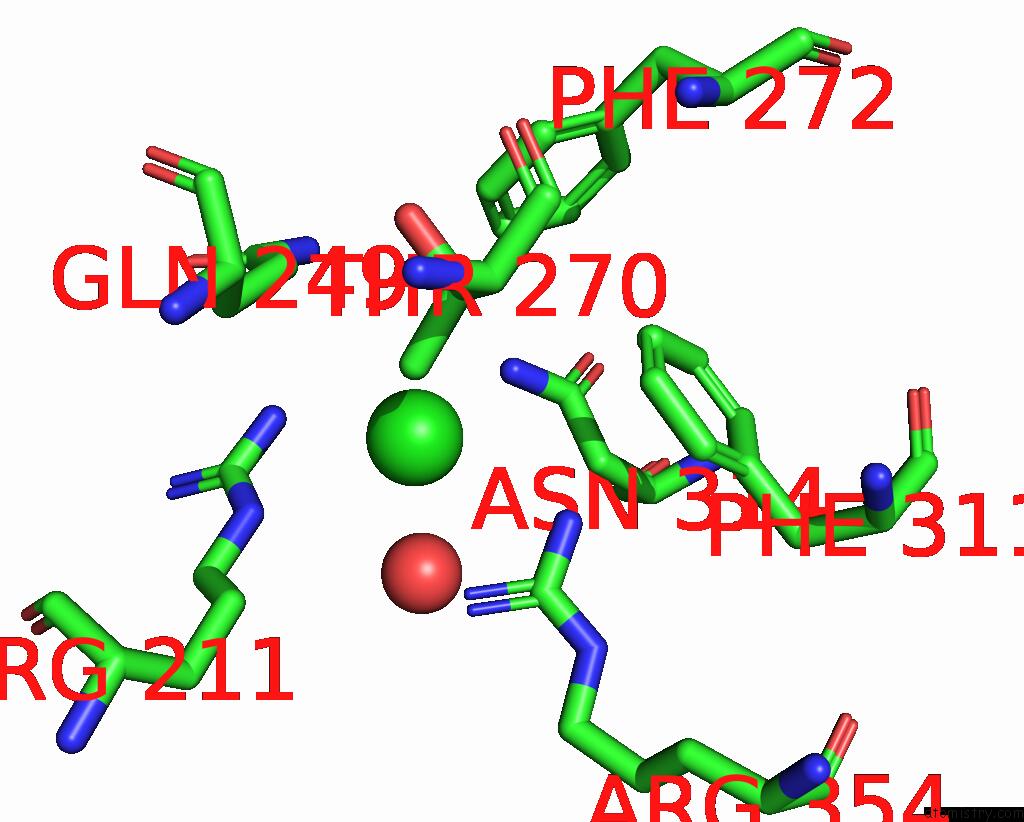
Mono view
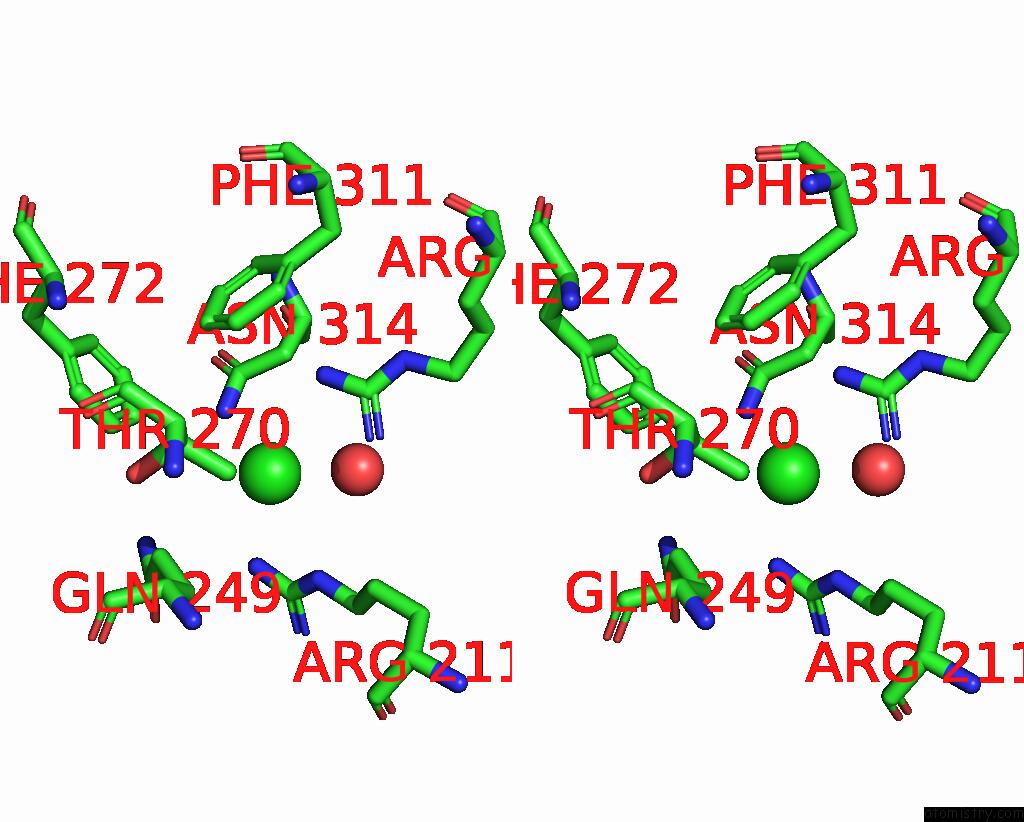
Stereo pair view
Mono view
Stereo pair view
A full contact list of Chlorine with other atoms in the Cl binding
site number 1 of X-Ray Crystal Structure of the E249Q Mutant of Alpha-Amylase I From Eisenia Fetida within 5.0Å range:
|
Reference:
Y.Hirano,
K.Tsukamoto,
S.Ariki,
Y.Naka,
M.Ueda,
T.Tamada.
X-Ray Crystallographic Structural Studies of Alpha-Amylase I From Eisenia Fetida. Acta Crystallogr D Struct V. 76 834 2020BIOL.
ISSN: ISSN 2059-7983
PubMed: 32876059
DOI: 10.1107/S2059798320010165
Page generated: Sun Jul 28 03:01:56 2024
ISSN: ISSN 2059-7983
PubMed: 32876059
DOI: 10.1107/S2059798320010165
Last articles
Zn in 9J0NZn in 9J0O
Zn in 9J0P
Zn in 9FJX
Zn in 9EKB
Zn in 9C0F
Zn in 9CAH
Zn in 9CH0
Zn in 9CH3
Zn in 9CH1