Chlorine »
PDB 6m83-6mj6 »
6mgb »
Chlorine in PDB 6mgb: Thermosulfurimonas Dismutans Kpsc, Beta Kdo 2,4 Transferase
Protein crystallography data
The structure of Thermosulfurimonas Dismutans Kpsc, Beta Kdo 2,4 Transferase, PDB code: 6mgb
was solved by
L.Doyle,
E.Mallette,
M.S.Kimber,
C.Whitfield,
with X-Ray Crystallography technique. A brief refinement statistics is given in the table below:
| Resolution Low / High (Å) | 46.23 / 1.80 |
| Space group | P 21 21 21 |
| Cell size a, b, c (Å), α, β, γ (°) | 50.020, 65.540, 121.080, 90.00, 90.00, 90.00 |
| R / Rfree (%) | 18.3 / 21.3 |
Other elements in 6mgb:
The structure of Thermosulfurimonas Dismutans Kpsc, Beta Kdo 2,4 Transferase also contains other interesting chemical elements:
| Magnesium | (Mg) | 1 atom |
Chlorine Binding Sites:
The binding sites of Chlorine atom in the Thermosulfurimonas Dismutans Kpsc, Beta Kdo 2,4 Transferase
(pdb code 6mgb). This binding sites where shown within
5.0 Angstroms radius around Chlorine atom.
In total only one binding site of Chlorine was determined in the Thermosulfurimonas Dismutans Kpsc, Beta Kdo 2,4 Transferase, PDB code: 6mgb:
In total only one binding site of Chlorine was determined in the Thermosulfurimonas Dismutans Kpsc, Beta Kdo 2,4 Transferase, PDB code: 6mgb:
Chlorine binding site 1 out of 1 in 6mgb
Go back to
Chlorine binding site 1 out
of 1 in the Thermosulfurimonas Dismutans Kpsc, Beta Kdo 2,4 Transferase
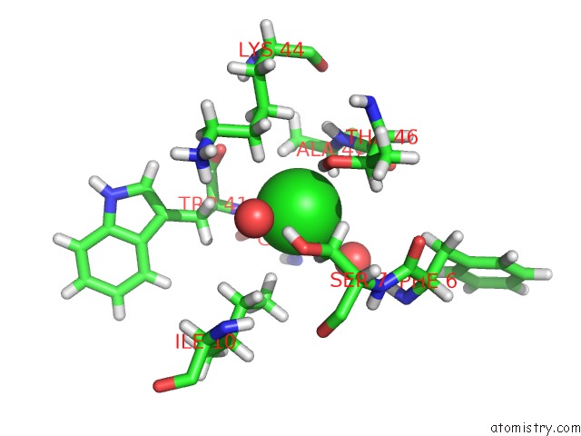
Mono view
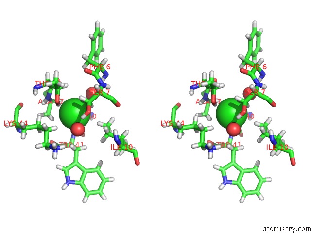
Stereo pair view
Mono view
Stereo pair view
A full contact list of Chlorine with other atoms in the Cl binding
site number 1 of Thermosulfurimonas Dismutans Kpsc, Beta Kdo 2,4 Transferase within 5.0Å range:
|
Reference:
L.Doyle,
O.G.Ovchinnikova,
K.Myler,
E.Mallette,
B.S.Huang,
T.L.Lowary,
M.S.Kimber,
C.Whitfield.
Biosynthesis of A Conserved Glycolipid Anchor For Gram-Negative Bacterial Capsules. Nat.Chem.Biol. V. 15 632 2019.
ISSN: ESSN 1552-4469
PubMed: 31036922
DOI: 10.1038/S41589-019-0276-8
Page generated: Sun Jul 28 03:12:32 2024
ISSN: ESSN 1552-4469
PubMed: 31036922
DOI: 10.1038/S41589-019-0276-8
Last articles
Zn in 9J0NZn in 9J0O
Zn in 9J0P
Zn in 9FJX
Zn in 9EKB
Zn in 9C0F
Zn in 9CAH
Zn in 9CH0
Zn in 9CH3
Zn in 9CH1