Chlorine »
PDB 6mjh-6mr3 »
6mo6 »
Chlorine in PDB 6mo6: Crystal Structure of the Selenomethionine-Substituted Human Sulfide:Quinone Oxidoreductase
Protein crystallography data
The structure of Crystal Structure of the Selenomethionine-Substituted Human Sulfide:Quinone Oxidoreductase, PDB code: 6mo6
was solved by
M.R.Jackson,
M.S.Jorns,
P.J.Loll,
with X-Ray Crystallography technique. A brief refinement statistics is given in the table below:
| Resolution Low / High (Å) | 62.69 / 2.59 |
| Space group | P 61 2 2 |
| Cell size a, b, c (Å), α, β, γ (°) | 119.390, 119.390, 551.858, 90.00, 90.00, 120.00 |
| R / Rfree (%) | 18.7 / 24.5 |
Chlorine Binding Sites:
The binding sites of Chlorine atom in the Crystal Structure of the Selenomethionine-Substituted Human Sulfide:Quinone Oxidoreductase
(pdb code 6mo6). This binding sites where shown within
5.0 Angstroms radius around Chlorine atom.
In total 4 binding sites of Chlorine where determined in the Crystal Structure of the Selenomethionine-Substituted Human Sulfide:Quinone Oxidoreductase, PDB code: 6mo6:
Jump to Chlorine binding site number: 1; 2; 3; 4;
In total 4 binding sites of Chlorine where determined in the Crystal Structure of the Selenomethionine-Substituted Human Sulfide:Quinone Oxidoreductase, PDB code: 6mo6:
Jump to Chlorine binding site number: 1; 2; 3; 4;
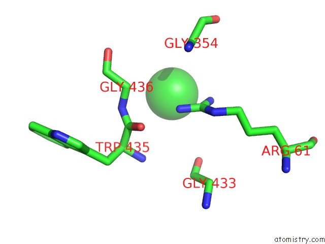
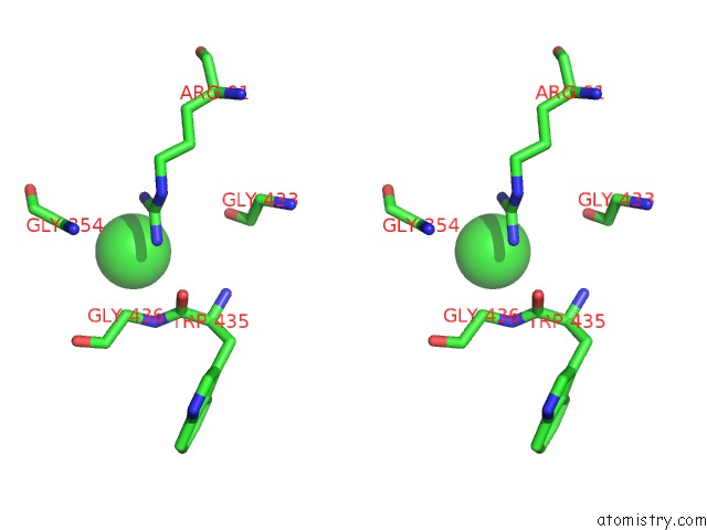
Chlorine binding site 1 out of 4 in 6mo6
Go back to
Chlorine binding site 1 out
of 4 in the Crystal Structure of the Selenomethionine-Substituted Human Sulfide:Quinone Oxidoreductase
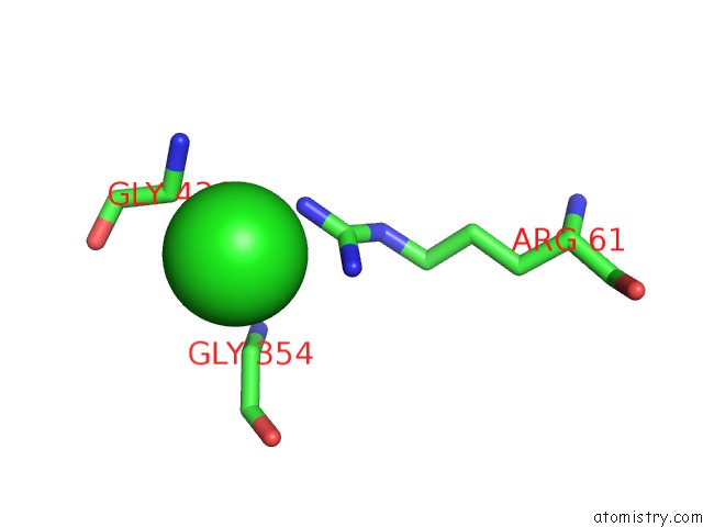
Mono view
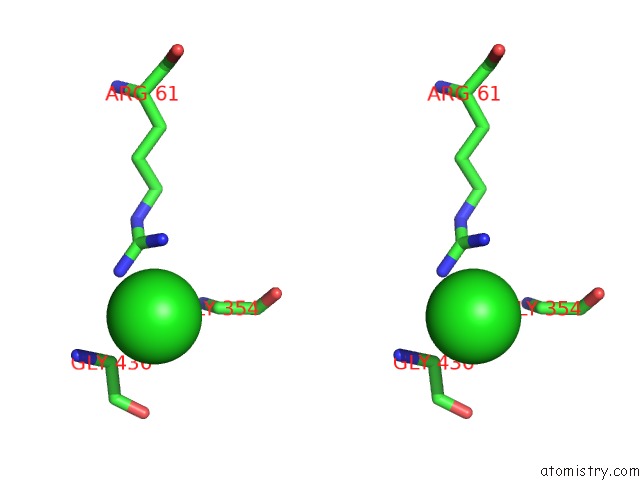
Stereo pair view
Mono view
Stereo pair view
A full contact list of Chlorine with other atoms in the Cl binding
site number 1 of Crystal Structure of the Selenomethionine-Substituted Human Sulfide:Quinone Oxidoreductase within 5.0Å range:
|
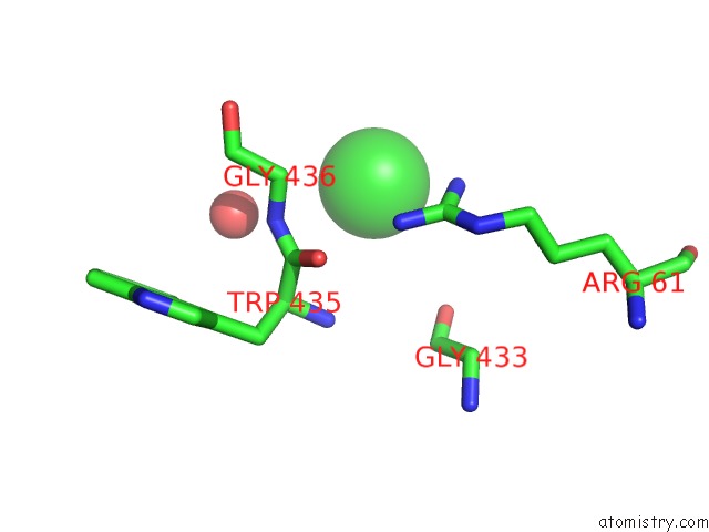
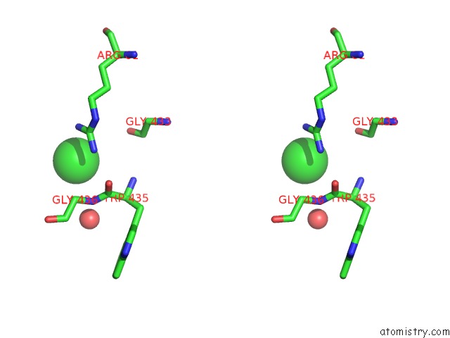
Chlorine binding site 2 out of 4 in 6mo6
Go back to
Chlorine binding site 2 out
of 4 in the Crystal Structure of the Selenomethionine-Substituted Human Sulfide:Quinone Oxidoreductase
Mono view
Stereo pair view
Mono view
Stereo pair view
A full contact list of Chlorine with other atoms in the Cl binding
site number 2 of Crystal Structure of the Selenomethionine-Substituted Human Sulfide:Quinone Oxidoreductase within 5.0Å range:
|
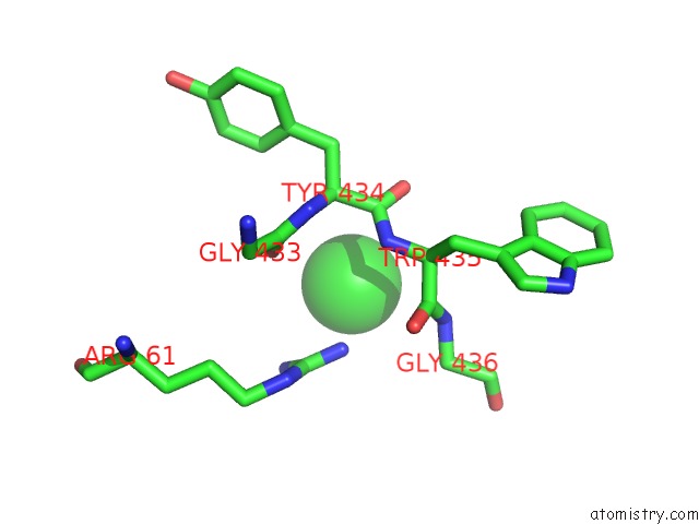
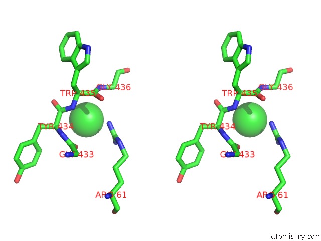
Chlorine binding site 3 out of 4 in 6mo6
Go back to
Chlorine binding site 3 out
of 4 in the Crystal Structure of the Selenomethionine-Substituted Human Sulfide:Quinone Oxidoreductase
Mono view
Stereo pair view
Mono view
Stereo pair view
A full contact list of Chlorine with other atoms in the Cl binding
site number 3 of Crystal Structure of the Selenomethionine-Substituted Human Sulfide:Quinone Oxidoreductase within 5.0Å range:
|
Chlorine binding site 4 out of 4 in 6mo6
Go back to
Chlorine binding site 4 out
of 4 in the Crystal Structure of the Selenomethionine-Substituted Human Sulfide:Quinone Oxidoreductase
Mono view
Stereo pair view
Mono view
Stereo pair view
A full contact list of Chlorine with other atoms in the Cl binding
site number 4 of Crystal Structure of the Selenomethionine-Substituted Human Sulfide:Quinone Oxidoreductase within 5.0Å range:
|
Reference:
M.R.Jackson,
P.J.Loll,
M.S.Jorns.
X-Ray Structure of Human Sulfide:Quinone Oxidoreductase: Insights Into the Mechanism of Mitochondrial Hydrogen Sulfide Oxidation. Structure V. 27 794 2019.
ISSN: ISSN 0969-2126
PubMed: 30905673
DOI: 10.1016/J.STR.2019.03.002
Page generated: Sun Jul 28 03:20:18 2024
ISSN: ISSN 0969-2126
PubMed: 30905673
DOI: 10.1016/J.STR.2019.03.002
Last articles
Zn in 9J0NZn in 9J0O
Zn in 9J0P
Zn in 9FJX
Zn in 9EKB
Zn in 9C0F
Zn in 9CAH
Zn in 9CH0
Zn in 9CH3
Zn in 9CH1