Chlorine »
PDB 6mjh-6mr3 »
6mqa »
Chlorine in PDB 6mqa: Structure of Hiv-1 Ca P207S
Protein crystallography data
The structure of Structure of Hiv-1 Ca P207S, PDB code: 6mqa
was solved by
S.S.Smaga,
Y.Xiong,
with X-Ray Crystallography technique. A brief refinement statistics is given in the table below:
| Resolution Low / High (Å) | 46.33 / 3.20 |
| Space group | P 6 |
| Cell size a, b, c (Å), α, β, γ (°) | 90.237, 90.237, 57.534, 90.00, 90.00, 120.00 |
| R / Rfree (%) | 18.6 / 21.9 |
Other elements in 6mqa:
The structure of Structure of Hiv-1 Ca P207S also contains other interesting chemical elements:
| Iodine | (I) | 3 atoms |
Chlorine Binding Sites:
The binding sites of Chlorine atom in the Structure of Hiv-1 Ca P207S
(pdb code 6mqa). This binding sites where shown within
5.0 Angstroms radius around Chlorine atom.
In total 2 binding sites of Chlorine where determined in the Structure of Hiv-1 Ca P207S, PDB code: 6mqa:
Jump to Chlorine binding site number: 1; 2;
In total 2 binding sites of Chlorine where determined in the Structure of Hiv-1 Ca P207S, PDB code: 6mqa:
Jump to Chlorine binding site number: 1; 2;
Chlorine binding site 1 out of 2 in 6mqa
Go back to
Chlorine binding site 1 out
of 2 in the Structure of Hiv-1 Ca P207S
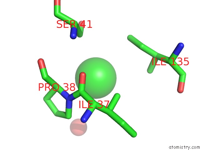
Mono view
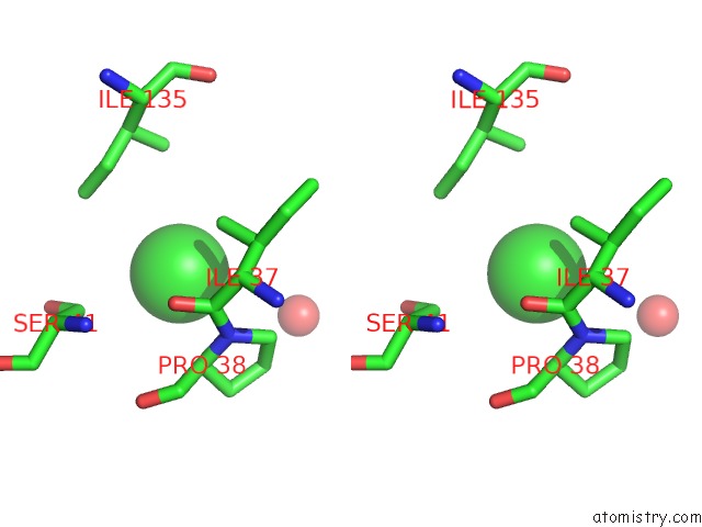
Stereo pair view
Mono view
Stereo pair view
A full contact list of Chlorine with other atoms in the Cl binding
site number 1 of Structure of Hiv-1 Ca P207S within 5.0Å range:
|
Chlorine binding site 2 out of 2 in 6mqa
Go back to
Chlorine binding site 2 out
of 2 in the Structure of Hiv-1 Ca P207S
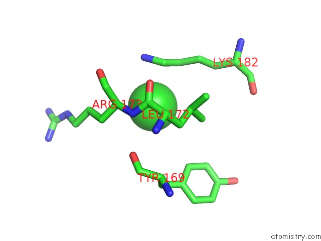
Mono view
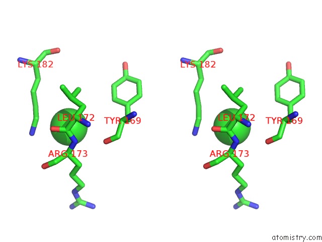
Stereo pair view
Mono view
Stereo pair view
A full contact list of Chlorine with other atoms in the Cl binding
site number 2 of Structure of Hiv-1 Ca P207S within 5.0Å range:
|
Reference:
S.S.Smaga,
C.Xu,
B.J.Summers,
K.M.Digianantonio,
J.R.Perilla,
Y.Xiong.
Mxb Restricts Hiv-1 By Targeting the Tri-Hexamer Interface of the Viral Capsid. Structure V. 27 1234 2019.
ISSN: ISSN 0969-2126
PubMed: 31155311
DOI: 10.1016/J.STR.2019.04.015
Page generated: Sun Jul 28 03:22:02 2024
ISSN: ISSN 0969-2126
PubMed: 31155311
DOI: 10.1016/J.STR.2019.04.015
Last articles
Zn in 9J0NZn in 9J0O
Zn in 9J0P
Zn in 9FJX
Zn in 9EKB
Zn in 9C0F
Zn in 9CAH
Zn in 9CH0
Zn in 9CH3
Zn in 9CH1